3D printing from cardiac CT data shows potential to aid TAVR
3D printing of models of the aortic root based on cardiac CT data sets may help reduce the risk of complications from transcatheter aortic valve replacement (TAVR). A proof-of-concept study conducted at Brigham and Women’s Hospital demonstrated that 3D aortic valve models could be accurately made from 2D data and could identify patients who would develop paravalvular aortic regurgitation (PAR) as a result of an incorrectly fitting prosthetic valve.
 Although the modeling techniques require additional development, the results of the feasibility study published in the January-February 2016 issue of the Journal of Cardiovascular Computed Tomography offer the hope that prosthetic valve fit may be improved. While TAVR is a safe alternative to surgery for some patients with aortic stenosis, prosthetic sizing and the complex 3D anatomy of the aortic root makes it difficult to predict how the prosthetic valve will adapt. Additionally if a circumferential seal is not achieved, PAR may occur.
Currently cardiac CT, transthoracic echocardiography (TTE), and/or transesophageal echocardiography (TEE) are used to determine fit and prosthetic valve selection. 3D advanced imaging processing visualization displayed on a two dimensional screen can show a patient’s root aortic root anatomy with great accuracy. However, the physical interactions of different types of prosthetic valves exert varying radial forces on the aortic root which cannot be inferred from 2D images and are difficult to model in a virtual 3D image-rendered space. Using a flexible 3D printed model may help to refine predictions of shape and or size mismatches by accounting for these physical interactions, lead author radiology resident Beth Ripley, M.D., and colleagues believe. For this reason, they wanted to determine if patient-specific 3D printed models could be used to visualize the fit between the native aortic valve complex and TAVR prosthetic valves, and also whether this information could predict the occurrence of post-procedural PAR.
The research team identified eight patients who experienced clinically documented PAR after TAVR and matched them by age, sex, and size of implanted valve with eight patients who did not have this complication. Two experienced cardiologists reviewed pre-TAVR cardiac CT and post-TAVR TTE. They determined that one of the control patients, actually had experienced mild PAR, and so the group for whom 3D models were made included 9 patients with PAR and 7 without.
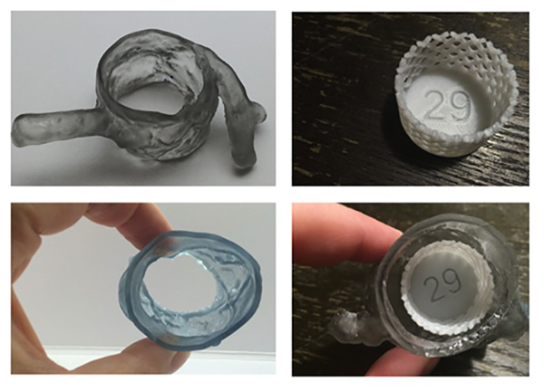
Multiphase data sets were reconstructed and segmented (excluding the left atrium and left ventricle. These segmented data sets were further processed. A sterolithography 3D printer created flexible aortic models, and hard plastic valve models were created on a material extrusion 3D printer. All models were compared with measurements made on cardiac CT images to verify accuracy of size.
To predict PAR, the authors positioned the valve model corresponding in size to each patient’s actual implanted prosthesis in the patient’s aortic model at the level of the aortic annulus. They photographed any light projecting through the left ventricular outflow tract, and used the findings to assess the presence of PAR.
The light transmission test correctly identified PAR in six of nine patients (66%) and correctly excluded PAR in five of seven patients (71%). The authors attributed lack of better accuracy to the fact that their current modeling technique to a variety of factors. However, they are optimistic that improvements will be made.
They wrote, “3D modeling presents a novel opportunity to plan TAVR device placement in situ. It may complement traditional techniques used to predict and potentially avoid complications such as PAR.”
REFERENCE
- Ripley B, Kelil T, Cheezum MK, et al. 3D printing based on cardiac CT assists anatomic visualization prior to transcatheter aortic valve replacement. 2016. J Cardiovasc Comput Tomogr. 10;1::28-36.
Citation
3D printing from cardiac CT data shows potential to aid TAVR. Appl Radiol.
March 23, 2016