3D Body Volume Scanner Uses AI to Predict Risk of Metabolic Syndrome
Images

Mayo Clinic researchers are using AI with an advanced 3D body-volume scanner to help doctors predict metabolic syndrome risk and severity. The combination of tools offers doctors a more precise alternative to other measures of disease risk like body mass index (BMI) and waist-to-hip ratio, according to findings published in the European Heart Journal - Digital Health.
Metabolic syndrome can lead to heart attack, stroke and other serious health issues and affects over a third of the US population and a quarter of people globally. The condition lacks widely accepted screening strategies. However, researchers found that using a 3D body volume scanner combined with imaging technology and Mayo Clinic-developed algorithms may help clinicians offer a more accurate method for identifying people who have the syndrome, as well as those at risk for developing it.
The effects of metabolic disease create hardship for patients. In addition to heart attack and stroke, people with metabolic syndrome are more likely to develop diabetes, cognitive disease and liver disease. Metabolic syndrome is diagnosed clinically when at least three of these five conditions are present: abdominal obesity, high blood pressure, high triglycerides, low HDL cholesterol and high fasting blood sugar.
"There is a need for a reliable, repeatable measure of metabolic syndrome risk and severity," says Betsy Medina Inojosa, MD, a research fellow at Mayo Clinic and first author of the study. "Body mass index measurements and bioimpedance scales that measure body fat and muscle are inaccurate for many people, and other types of scans are not widely available. Our research shows that this AI model may also be a tool to guide clinicians and patients to take action and seek outcomes that are a better fit for their metabolic health."
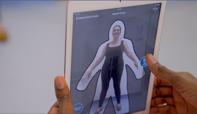
To develop the tool, researchers trained and validated an AI model on 1,280 volunteer subjects who underwent an evaluation that included 3D body-volume scans, standardized clinical questionnaires, blood tests and traditional body shape measurements. An extra 133 volunteers had front- and side-view images taken via a mobile app from Select Research called myBVI to further test the tool's ability to evaluate whether they had metabolic syndrome, and if so, how severe it was.
People with metabolic syndrome typically have apple-shaped bodies, meaning they carry a lot of their weight around the abdomen. The diagnosis of metabolic syndrome revolves around laboratory tests, blood pressure and body shape measurements, but there are no widely accepted routine screening strategies because these measurements are not always available or reproducible in the same way.
"This small study finds that digitally measuring a patient's body volume index with 3D imaging provides a highly accurate measurement of shapes and volumes in critical regions where unhealthy visceral fat is deposited, such as the abdomen and chest," says Francisco Lopez-Jimenez, MD, director of Preventive Cardiology at Mayo Clinic in Rochester and senior author of the study. "The scans also record the volume of hips, buttocks and legs – a measure related to muscle mass and 'healthy' fat. The 3D information about body volume in these key regions, whether from the large, stationary 3D scanner or from the mobile app, accurately flagged the presence and severity of metabolic syndrome using imaging instead of invasive tests. Looking ahead, the next steps will be to broaden the sample of research subjects to include more diversity."
Related Articles
Citation
. 3D Body Volume Scanner Uses AI to Predict Risk of Metabolic Syndrome. Appl Radiol.
August 27, 2024