Contrast-Enhanced In-Phase Dixon Sequence for Detecting Breast Biopsy Clips
 Utilizing a contrast-enhanced in-phase Dixon sequence identifies areas of prior biopsy during breast MRI interpretation and reduces the need to correlate with other imaging methods. The study, published in the American Journal of Roentgenology reports that employing this sequence may “help address a current challenge in routine clinical breast MRI interpretation,” according to the authors.
“Compared with clinical sequences, contrast-enhanced in-phase Dixon had higher sensitivity for detecting breast biopsy clips on MRI, as well as higher reader confidence and contrast-to-noise ratio (CNR), without change in positive predictive value (PPV),” wrote corresponding and co-first author, Michael W. Taylor-Cho, MD, MPH, of Duke University Medical Center in Durham, NC.
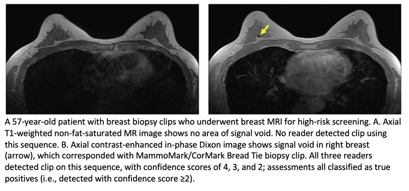
Dr Taylor-Cho and team’s retrospective study numbered 164 women (mean age, 50.3 years) with a total of 281 breast biopsy clips who underwent contrast-enhanced breast MRI between January 2, 2019, and April 16, 2020. Blinded to patient identifiers, sequence information, and biopsy clip details, three radiologists independently annotated their findings on three clinical sequences—T1-weighted (T1W) non-fat-suppressed (NFS), STIR, first phase from dynamic contrast-enhanced T1W fat-suppressed (FS) —as well as contrast-enhanced in-phase Dixon. Confidence was then recorded on a scale of 1–4.
Ultimately, compared with T1W NSF, STIR, and T1WFS sequences, the contrast-enhanced in-phase Dixon sequence evidenced the highest sensitivity for breast biopsy clip detection (85.1% vs. 26.6%-78.2%), highest reader confidence (3.5 vs. 1.7–3.0), and highest CNR (4.05 vs 0.54–1.21), without a significant difference in PPV (96.4% vs. 92.2%–96.1%).