Wireless Wearable Ultrasound Monitors Muscle Activity
Images

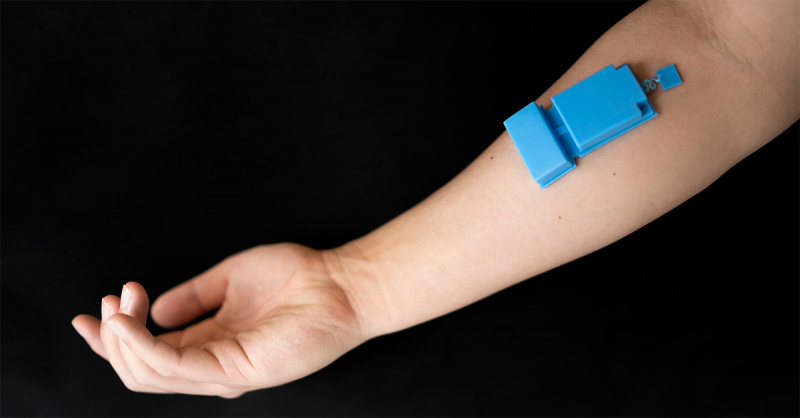
Engineers at the University of California San Diego have developed a wearable ultrasound device that can provide long-term, wireless monitoring of muscle activity with potential applications in healthcare and human-machine interfaces. Designed to stick to the skin with a layer of adhesive and powered by a battery, the device enables high-resolution tracking of muscle function without invasive procedures.
A team of researchers led by Sheng Xu, a professor and Jacobs Faculty Scholar in the Aiiso Yufeng Li Family Department of Chemical and Nano Engineering at UC San Diego, published their work in Nature Electronics. The work was a collaborative project with Jinghong Li, a pulmonologist, intensive care specialist and professor of medicine at UC San Diego Health.
In tests, the device was worn over the rib cage to monitor diaphragm motion and thickness, which are useful for assessing respiratory health. “By tracking diaphragm activity, the technology could potentially support patients with respiratory conditions and those reliant on mechanical ventilation,” said Joseph Wang, a distinguished professor in the Aiiso Yufeng Li Family Department of Chemical and Nano Engineering who is a co-author on the study.
Additionally, researchers successfully used the device on the forearm to capture hand and wrist muscle activity, which enabled its use as a human-machine interface to control a robotic arm and play a virtual game.
This wearable ultrasound technology may offer a promising new alternative to the current clinical standard, electromyography (EMG), which involves applying metal electrodes on the skin to record electrical muscle activity. Despite EMG’s longstanding use, it suffers from low resolution and weak signals. For example, signals from multiple muscle fibers often blend together, making it challenging to isolate the contributions of specific muscle fibers.
Ultrasound, however, provides high-resolution imaging by penetrating deep tissues, offering detailed insights into muscle function. The ultrasound technology that Xu’s team and their collaborators developed has the additional advantages of being compact, wireless, and low-power. “This technology could potentially be worn by individuals during their daily routines for continuous, long-term monitoring,” said study co-first author Xiangjun Chen, a PhD candidate in the Materials Science and Engineering program at UC San Diego.
The device is housed in a flexible silicone elastomer casing and consists of three main components: a single transducer for sending and receiving ultrasound waves; a custom-designed wireless circuit that controls the transducer, records data and wirelessly transmits the data to a computer; and a lithium-polymer battery that can power the system for at least three hours.
A key innovation of this work is the use of a single ultrasound transducer to sense deep tissues effectively. The transducer emits intensity-controlled ultrasound waves and captures radiofrequency signals that carry rich information, enabling clinical applications such as measuring diaphragm thickness. Using these signals, the device can achieve high spatial resolution, which is key for isolating specific muscle movements. To extract additional insights from these signals, the researchers developed an artificial intelligence algorithm that maps the signals to their corresponding muscle distributions, enabling it to identify specific hand gestures from the collected signals with high accuracy and reliability.
When worn on the rib cage, the device can accurately monitor diaphragm thickness with submillimeter precision. Diaphragm thickness is a metric used in the clinic to evaluate diaphragm dysfunction and predict outcomes in ventilated patients. By analyzing muscle motion, the researchers could also detect different breathing patterns, such as shallow and deep breaths. This functionality could help diagnose conditions linked to breathing irregularities, such as asthma, pneumonia and chronic obstructive pulmonary disease (COPD). In a small group trial, the device successfully distinguished breathing patterns of individuals with COPD from those of healthy participants.
“This demonstrates the technology’s potential for clinical applications in respiratory care,” said study co-first author Muyang Lin, a postdoctoral researcher in the Aiiso Yufeng Li Family Department of Chemical and Nano Engineering at UC San Diego.
When worn on the forearm, the device offers precise tracking of muscle motion in the hands and wrists. Thanks to the artificial intelligence algorithm that the team developed, the system is capable of recognizing various hand gestures solely from the ultrasound signals. The system is able to recognize 13 degrees of freedom, covering 10 finger joints and three rotation angles of the wrist. As a result, it can capture even slight wrist and finger movements with high sensitivity.
In proof-of-concept tests, participants used the device on their forearms to control a robotic arm to pipette water into beakers. In another demonstration, they used the device to play a virtual game, using wrist movements to control a virtual bird’s flight through obstacles. “These demonstrations underscore the technology’s potential for prosthetics, gaming and other human-machine interface applications,” said study co-first author Wentong Yue, a PhD candidate in the Aiiso Yufeng Li Family Department of Chemical and Nano Engineering at UC San Diego.
Moving forward, the researchers plan to improve the technology’s accuracy, portability, energy efficiency and computational capabilities.
Related Articles
Citation
Wireless Wearable Ultrasound Monitors Muscle Activity. Appl Radiol.
November 3, 2024