Study Confirms Sub-Millimeter Precision for XPlan.AI’s X-ray-Based 3D Bone Modeling System
Images
XPlan.ai announced the publication of a peer-reviewed clinical study confirming, sub-mm accuracy in a variety of clinically relevant measurements for its X-ray based 3D bone modeling system. The study, led by a consortium of orthopedic surgeons and published in the Journal of Clinical Medicine, found that XPlan.ai offers a promising alternative to conventional CT scans, opening up the market for image-based computer-assisted surgery such as robotics, AR, and navigation while increasing efficiency, saving costs and avoiding unnecessary radiation exposure.
XPlan.ai uses advanced artificial intelligence (AI) to produce accurate 3-dimensional bone models from two standard X-ray images. Together with XPlan's automated planning technologies, this model can be used for surgical planning and navigation during orthopedic procedures such as total knee replacement, potentially providing incredibly large patient populations with the most advanced care while avoiding the cost, time, administrative overhead, reimbursement issues, and added radiation involved in a conventional CT scan.
"Replacing CT scans with standard, universally available X-rays has long been considered a 'holy grail' of computer-assisted orthopedic surgery," said Moshe Safran, CEO at XPlan.ai. "Accuracy and robustness have been the key challenges, and our technology provides unique capabilities in this regard, paving the way for universal access to image-based computer assisted surgery."
XPlan.ai 's groundbreaking technology provides two main benefits: clinical and operational. The clinical benefit is lower levels of radiation exposure from X-ray imaging compared to a complete knee CT scan. Operationally, all relevant patients routinely undergo X-ray imaging, offering much better accessibility than CT. Additionally, X-rays are more widely reimbursed in the US healthcare system, are often lower in cost, and offer a quicker and more streamlined patient journey from diagnosis to the OR.
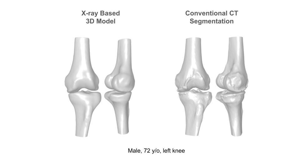
The clinical evaluation of this tool was conducted using imaging from total knee replacement patients from Assuta Medical Center in Tel-Aviv, a leading medical center in Israel. Unlike cadaver-based studies often used in the orthopedic technology space, the patients enrolled in this study had pathological anatomies typical of real-world clinical cases. The accuracy of the tool was proven by comparing the resulting 3D models to the ground truth patient anatomy given in a corresponding CT scan. The accuracy was measured in multiple areas that are used for actual surgical planning, including bony landmarks and anatomical axes, and was found to be equivalent to CT-based measurements at a sub-mm level across the board.
"Today's orthopedic patients demand precise and personalized care, incorporating technologies such as AR and surgical robotics. Using a 3D image-based preoperative model to plan the case is the best approach, enabling surgeons to be better prepared and saving precious time in the OR," said Dr Vadim Benkovitch, Head of Orthopedic Department at University Medical Center Soroka and Founder & Medical Director of the Israeli Joint Health Center at Assuta Medical Center. "This is a win for both patients and providers - both reducing the chances of a complication or infection, and at the same time improving efficiency and providing care to more patients with the same amount of resources. I am encouraged to see that the accuracy of XPlan's solution has passed the most stringent tests conducted in our study."
Going forward, XPlan.ai plans to apply for FDA clearance of its knee reconstruction solution. In parallel, further applications are under development for additional anatomies, with promising initial results indicating wide applicability of XPlan's unique technology.
Related Articles
Citation
Study Confirms Sub-Millimeter Precision for XPlan.AI’s X-ray-Based 3D Bone Modeling System. Appl Radiol.
March 25, 2024