State-of-the-Art Mammography in 2007
Images


Dr. Hixson is a Women's Imaging Specialist in Chattanooga, TN and the founder of American Mammographics, Inc.
If a facility has not changed the way it performs mammography in the last five years, its work probably is not state-of-the-art. Too many cancers are still being missed, especially in patients with denser breasts. Higher contrast resolution is needed. This can be achieved with either full-field digital mammography (FFDM) or one of the new film-screen mammography (FSM) systems that utilize double emulsion film.
Digital vs. Film-Screen Mammography
Is FFDM necessary in order to perform state-of-the-art mammography? There have been four screening trials comparing FFDM and FSM. None of these trials has shown a significant difference in the diagnostic accuracy between the two in the population as a whole. The latest trial was the ACRIN study (DMIST), which compared FFDM with FSM in 42,760 women. 1 DMIST initially reported that FFDM showed some increased sensitivity in heterogeneously dense or extremely dense breasts (mainly for DCIS). However, subsequent analysis of the detected cancers failed to show any difference in sensitivity even for denser breasts. 2
Awareness of the limitations of DMIST and the other three trials will allow more meaningful scrutiny of their results and conclusions. The various types of bias and limitations in scientific investigations have been comprehensively described. 3 When a new test is evaluated, its efficacy is determined by comparing its performance to that of the accepted reference test. A single emulsion film system was the reference test with which digital was compared in all of these trials. However, FSM technology has subsequently been significantly improved. The new double emulsion film-screen systems have now become the reference test with which digital must be compared. The results of DMIST and the other trials are not valid for comparing the performance of FFDM with the new generation of FSM systems, which use double emulsion film technology.
S.O.F.T. Paddle ®
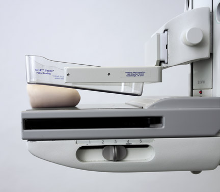
The S.O.F.T. Paddle ® is one of several recently developed products that can enhance the performance of FSM. A conventional compression paddle produces optimum compression of only the thick posterior portion of the breast. Several compression paddles have been developed that rotate as compression is applied. They tilt downward over the anterior breast, but at the same time, they rotate upward posteriorly. Unfortunately, this causes suboptimal compression of the posterior breast, which is where most cancers occur.
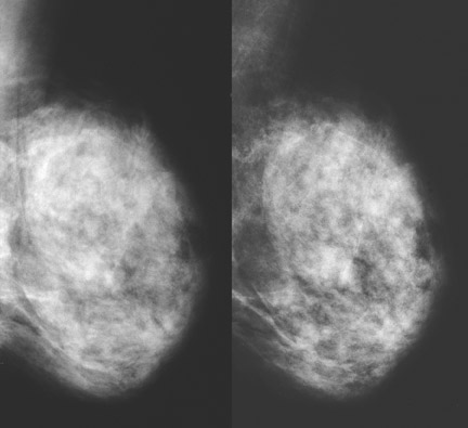
The S.O.F.T. Paddle ® from American Mammographics is different (Figure 1). The compression surface is horizontal near the chest wall, then curves slightly to slope downward toward the nipple. It does not rotate. Increased compression of the anterior two thirds of the breast is achieved while maintaining excellent compression posteriorly. Fibroglandular tissue is spread apart more effectively so that a cancer is less likely to be obscured by superimposed structures. There is additional reduction in breast thickness. This permits a lower kV, which increases contrast. Exposure time is also shortened, which eliminates motion blur. The improved image quality with S.O.F.T. Paddle ® is shown in Figure 2. S.O.F.T. Paddle ® provides many potential benefits, such as increased sensitivity for cancer detection and fewer callbacks (Table 1).
Dr. László Tabár says of the S.O.F.T. Paddle ® , "the image details that are so essential in making the first and most important decision (callback or normal) are visualized so well and with such sharpness as I have never experienced it previously." 4
Improved Film-Screen Technology
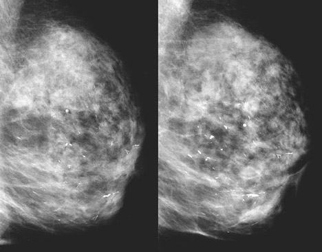
Newer state-of-the-art FSM systems, such as Kodak Min-R EV, utilize double emulsion film with smaller, more uniform cubic silver halide grains that increase contrast and image sharpness. This will increase detection of cancers in denser breasts. Dramatic improvement in image quality occurs when a newer FSM system and S.O.F.T. Paddle ® are used together (Figure 3).
Optimized Techniques
Since cancers are most likely to be missed in dense breasts, better penetrating, higher energy radiation should now be used routinely for all screening mammography. kV could be increased, but this would reduce contrast. To produce the higher energy radiation, it is better to substitute the rhodium (Rh) filter for the molybdenum (Mo) filter. This will result in a more penetrating beam and will shorten the exposure.
An important study in Sweden showed that the Rh filter provided better depiction of glandular tissue and the skin for breasts of all size and glandular composition, while giving a lower dose. 5 Hendrick et al 6 showed that when the Rh filter is used for breasts ≥6 cm in thickness, the exposure time is reduced by as much as 30%. The resulting shorter exposure will reduce motion blur. It will also permit the use of a lower kV, which will increase contrast. The Rh filter also reduces glandular dose by ≥20% in breasts ≥6 cm in thickness.
Pros and Cons of Digital Mammography
The primary motivation for switching to FFDM is not necessarily the superiority of detector technology but rather the intrinsic value of the digital image format. This permits images to be manipulated and transmitted via teleradiology. Patient throughput also is faster, which increases technologist productivity. In an editorial accompanying the DMIST article, Dershaw 7 recognized several benefits of FFDM but said that these advantages must be weighed against the cost of FFDM systems (up to five times as expensive as FSM systems). He also stated that "more time and effort are often required to read digital mammograms than film mammograms." 7 A study by Berns et al 8 showed that digital mammography reduced the time needed to image a patient by 35% but increased interpretation time by 57%. Barbara Monsees, MD, Mallinckrodt Institute of Radiology, said, "Every single one of us at Mallinckrodt was surprised by how much longer it takes to interpret digital images…I think time needs to be factored into any cost-effectiveness analysis. This could become a workforce issue." 9
Summary
State-of-the-art mammography can be achieved with either digital or film-screen mammography. Film-screen mammography has been significantly improved by new products that were not available at the time DMIST and the three other trials were conducted. New generation double emulsion film-screen technology produces increased image contrast and sharpness. Better breast compression can be achieved with the unique S.O.F.T. Paddle ® (Figure 2). Greater use of the Rh filter will generate higher energy radiation for better penetration and reduced radiation dose. Patients, providers, and third-party payers can be confident that digital and state-of-the-art film-screen mammography are equally effective for improving the detection of cancer of the breast.