Spontaneous ureteral injury
Images


CASE SUMMARY
An 18-year-old gravida 3 woman was transferred from an outside hospital to our facility for worsening abdominal pain, tachycardia, and mild hypotension following a reportedly uncomplicated vaginal delivery. The outside hospital CT scan demonstrated a large amount of free fluid measuring simple fluid attenuation within the right pelvis. Upon arrival, the patient’s symptoms worsened and another CT scan was obtained with a split bolus technique1 to capture the nephrographic and excretory phases in combination, which demonstrated right ureteral injury with extravasation of urine within the pelvis. The patient was then taken to urgent cystoscopy for retrograde ureteral stent placement, which was unsuccessful. Interventional radiology subsequently placed a percutaneous nephroureteral stent past the point of injury and into the bladder, which was successfully internalized to a double J ureteral catheter 2 weeks later.
IMAGING FINDINGS
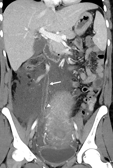
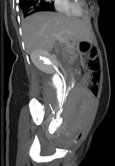
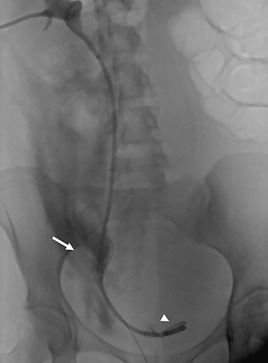
Initial CT scan from the outside hospital demonstrated a dilated, enhancing right ureter terminating in a large amount of simple fluid within the pelvis (Figure 1), with a fleck of gas adjacent to the distal ureter. A Foley catheter and gas were also seen in the bladder lumen. Subsequent CT at our institution was performed with a split bolus technique to allow for opacification of the collecting systems. This study demonstrated contrast extravasation from the injured distal right ureter (Figure 2). Fluoroscopic images from placement of a percutaneous nephroureteral stent confirmed the ureteral injury, with free contrast accumulating adjacent to the wire during placement and the successful placement of a percutaneous nephroureteral stent crossing the site of injury to terminate in the bladder (Figure 3).
DIAGNOSIS
Spontaneous distal ureteral injury following uncomplicated vaginal delivery
DISCUSSION
Spontaneous ureteral injury, defined as injury in the absence of interventional procedure, surgery, or trauma, is a rarely reported cause of acute abdominal pain.2-4 Most commonly due to ureteral stones, they typically occur in the proximal ureter.2-4 Ureteral rupture during pregnancy is thought to result from increased pressure secondary to ureteral compression.2,5 Vaginal delivery is an even more rarely documented cause of spontaneous ureteral injury. In our case, it was presumably due to the downward pressure placed on the bladder during active labor, placing the ureter in traction. Spontaneous ureteral injury can present with symptoms similar to other entities found during pregnancy and/or immediately postpartum, such as appendicitis, pyelonephritis, renal calculi, and uterine rupture.2,3,5 A CT scan using split bolus technique that captures information both in the normal portal venous and excretory phases may be valuable in narrowing the differential diagnosis.
In a postpartum patient with a history of uncomplicated vaginal delivery, the presence of a large amount of simple free fluid immediately places the urinary tract injury as the most probable source. In our case, the presence of bladder gas and gas adjacent to an abnormal right ureter deepened suspicion of a ureteral injury. When appropriately performed, a contrast-enhanced CT containing information in the excretory phase can easily confirm this diagnosis and potentially delineate the level of injury for treatment planning. The key is to recognize large-volume, simple free fluid in this rare clinical scenario as a trigger for repeat, delayed scanning in the excretory phase. The patient could then proceed to surgery, urgent cystoscopy for stent placement, or be treated with conservative management.3
CONCLUSION
Spontaneous ureteral injury is a rare complication of vaginal delivery, but it should be strongly suspected when a large amount of simple free fluid is seen on CT. Delayed phase CT, either excretory phase or combined nephrographic/excretory phase, can further characterize the injury for treatment planning. Many cases of ureteral injury can be managed with a relatively conservative approach, including ureteral stenting, without requiring open surgical repair.3,4
REFERENCES
- Chow LC, Kwan SW, Olcott EW, Sommer G. Split-bolus MDCT urography with synchronous nephrographic and excretory phase enhancement. AJR Am J Roentgenol. 2007;189(2):314-322.
- Narasimhulu DM, Egbert NM, Matthew S. Intrapartum spontaneous ureteral rupture. Obstet Gynecol. 2015;126(3):610-612.
- Chen GH, Hsiao PJ, Chang YH, Chen CC, Wu HC, Yang CR, et al. Spontaneous ureteral rupture and review of the literature. Am J Emerg Med. 2014;32(7):772-774.
- Stravodimos K, Adamakis I, Koutalellis G, Koritsiadis G, Grigoriou I, Screpetis K, et al. Spontaneous perforation of the ureter: Clinical presentation and endourologic management. J Endourol. 2008;22(3):479-484.
- Upputalla R, Moore RM, Jim B. Spontaneous forniceal rupture in pregnancy. Case Rep Nephrol. 2015;2015:379061.
Citation
KL R, M L, S B,. Spontaneous ureteral injury. Appl Radiol. 2018;(7):38-39.
July 6, 2018