SNMMI Image of the Year: Ultra-High-Performance PET Delivers Never-Before-Seen Images of the Brain Nuclei
Images
A new ultra-high-performance brain PET system, NeuroEXPLORER, allows for the direct measurement of brain nuclei as never before seen or quantified with its ultra-high sensitivity and resolution. The NeuroEXPLORER provides exceptional brain PET images and has the potential to spur advances in the treatment of many brain diseases. This research was presented at the 2024 Society of Nuclear Medicine and Molecular Imaging (SNMMI) Annual Meeting, and the grouping of images highlighting targeted tracer uptake in specific brain nuclei has been selected as the 2024 SNMMI Henry N. Wagner, Jr., Image of the Year.
Each year, SNMMI chooses an image that best exemplifies the most promising advances in the field of nuclear medicine and molecular imaging. The state-of-the-art technologies captured in these images demonstrate the capacity to improve patient care by detecting disease, aiding diagnosis, improving clinical confidence, and providing a means of selecting appropriate treatments. This year, the SNMMI Image of the Year was chosen from more than 1,500 abstracts submitted for the meeting.
The image quality of PET systems has improved in recent years, mostly by increases in sensitivity, including enhanced time-of-flight capabilities. However, these systems have shown only minimal improvement in intrinsic resolution. To address these issues, researchers designed the NeuroEXPLORER PET scanner with a focus on ultra-high sensitivity and resolution, as well as continuous head motion correction.
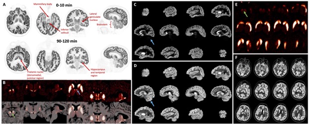
In the study, researchers conducted human brain imaging with both the NeuroEXPLORER and the High Resolution Research Tomograph, or HRRT (the previous state-of-the-art imaging tool). Multiple targeted radiopharmaceuticals were administered to observe synaptic density, dopamine receptors and transporters, muscarinic cholinergic receptors, and glutamate receptors. Images from both scanners were then compared.
A striking improvement in image contrast and quality of the NeuroEXPLORER compared to the HRRT was evident. NeuroEXPLORER images demonstrated low noise and exquisite resolution, showing focal uptake in specific brain nuclei.
“The high resolution of NeuroEXPLORER images is due to the system’s unique detector design and exceptional sensitivity produced by its long axial field-of-view,” said Richard E. Carson, PhD, professor of Biomedical Engineering and of Radiology and Biomedical Imaging and Emeritus director of the PET Center at Yale University in New Haven, Connecticut. “This technology will provide the opportunity for advanced research on all types of neuronal molecular and functional activity.”
“The dramatic improvement in resolution and overall quality of the NeuroEXPLORER images compared to the HRRT images is clear,” noted SNMMI Scientific Program Committee chair Heather Jacene, MD. “The NeuroEXPLORER has the potential to be a gamechanger in research for conditions such as Alzheimer disease, Parkinson disease, epilepsy, and mental illnesses.”
The NeuroEXPLORER scanner was built as a collaboration with Yale University, University of California, Davis, and United Imaging Healthcare of America, and was funded by a National Institutes of Health Brain Initiative grant. While the NeuroEXPLORER is currently used for research purposes, Carson and colleagues hope that once the excellent image quality is recognized by physicians it will become available for clinical use.