Siemens Launches Wide-Angle 3D Mammography System
Images
Siemens Healthineers has launched Mammomat B.brilliant1, a new tomosynthesis mammography system with a wide-angle tube that moves around the breast at a 50° angle and a scan time of around five seconds, creating a 3D image with high depth resolution in the shortest time possible. The system enables abnormalities and microcalcifications in the tissue to be identified with a high level of accuracy. According to the company, the acquisition speed is about 35% faster compared to other devices, making the examination more comfortable for patients.
"Our systems have to provide meaningful answers in the shortest possible time. This is especially important in mammography, when the stressful uncertainty for patients must be reduced to a minimum. Only Mammomat B.brilliant offers this unique combination of speed, resolution and wide angle," says Verena Schön, Head of X-Ray Products at Siemens Healthineers.
"We are already benefiting from the significant benefits that Mammomat B.brilliant offers in our clinical routines. The speed of tomosynthesis is very impressive and the image quality is excellent. Together with the quick and convenient positioning of the patients, it is an excellent tool for us in mammography," says Dr. Machteld Keupers, UZ Leuven, Belgium.
Patient information and work steps are clearly visible from every angle on a prominent display monitor. The new ComfortMove function relieves users of physical strain, ensuring that physicians can easily and quickly adapt the device to the patient's anatomy. ComfortMove, the display and a positioning laser help to significantly accelerate scan preparation.
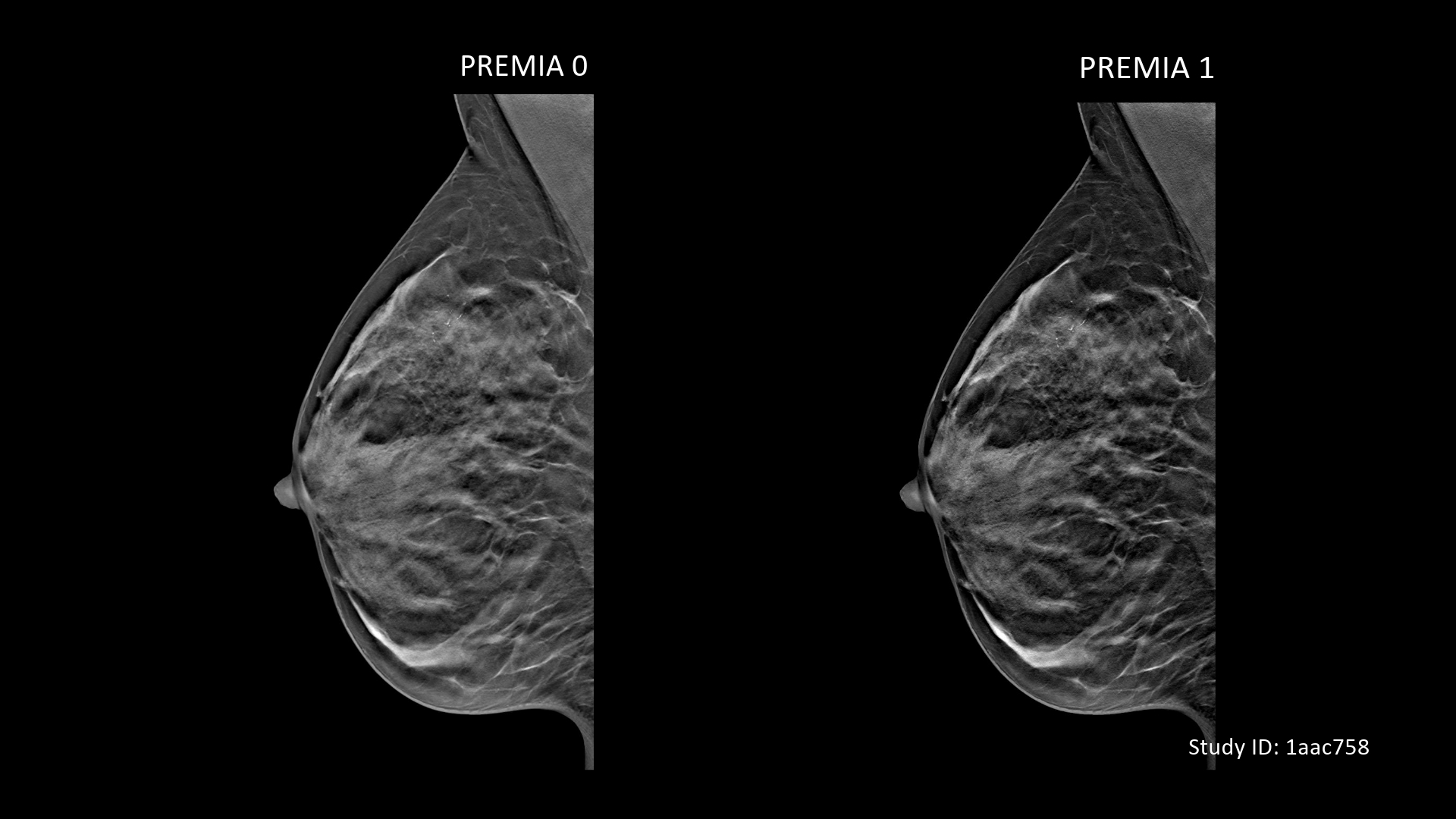
Mammomat B.brilliant is the first device from Siemens Healthineers with PlatinumTomo: the combination of wide-angle tomosynthesis, the new detector, the completely newly developed Flying Focus Spot tube and the Premia AI reconstruction to improve image quality. Flying focal spot technology is used for the first time in mammography: It prevents blurring effects caused by tube movements during tomosynthesis. This results in sharp images even at very high scanning speeds. In tomosynthesis, the X-ray tube moves in a circle around the breast, unlike conventional mammography, where it remains static – creating a 3D image.