Revolutionizing Brain MRI Acquisition
Images

Magnetic resonance imaging has seen significant strides in automation, with innovative solutions such as autoadaptive protocoling leading the way. These technologies are reshaping brain MRI protocols, offering unparalleled precision and expediting the diagnostic process.
The Evolution of MRI Protocols
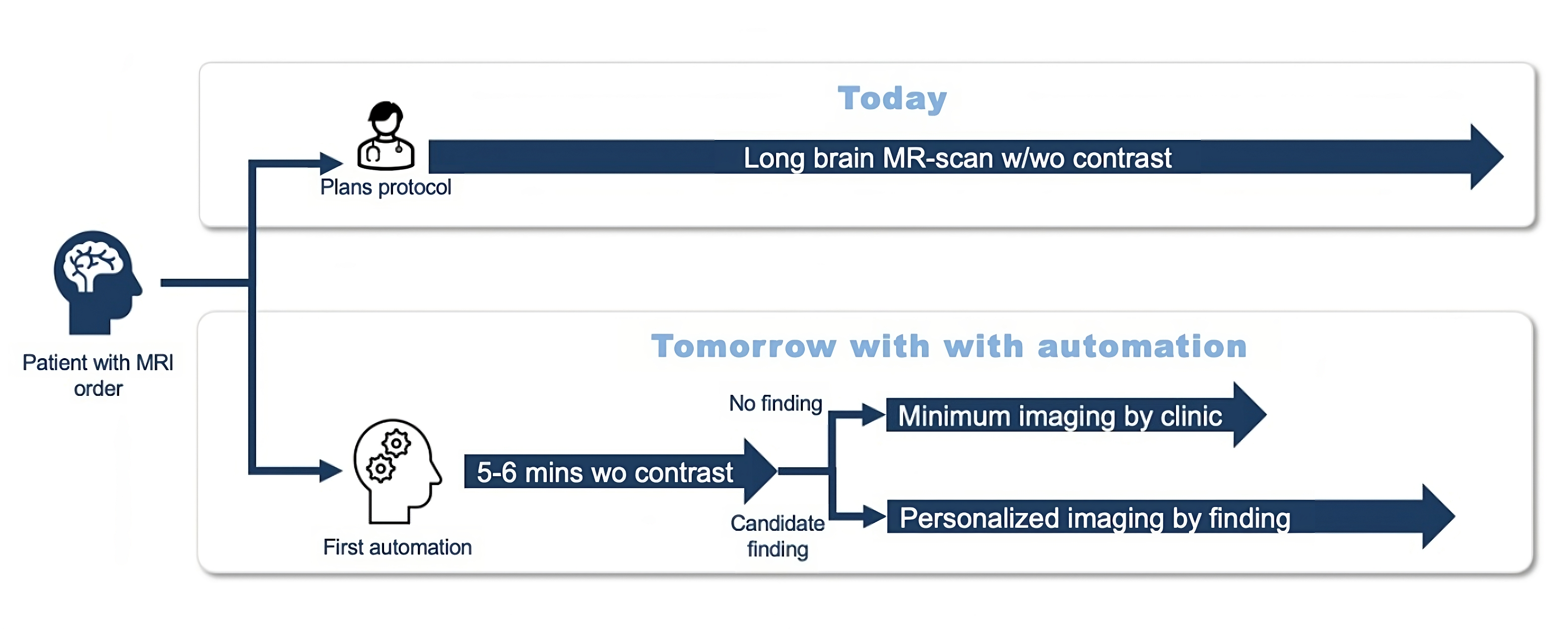
Traditionally, MRI protocoling has been largely a manual operation, requiring radiologists to meticulously configure imaging for each patient based on clinical indications. This process, while effective, can be time consuming and prone to human error.1,2 In response to these challenges, the industry has made a paradigm shift towards automation, aiming to streamline workflows, improve consistency, and ultimately deliver better patient outcomes.3
Advances in Protocol Optimization
Emerging artificial intelligence (AI)-based solutions have the potential to play a significant role in automating of brain MRI protocols. These technologies utilize AI and machine learning algorithms to sift through extensive datasets and derive imaging protocols customized to the individual characteristics of each patient. By analyzing a few initial sequences such as FLAIR, DWI, and others (eg, T2*, GRE, SWI), these systems can recommend the most suitable scanning sequences based on preliminary detection of conditions like tumors (for contrast) or infarctions (for vascular imaging).
This approach not only refines the personalization of patient care but also converts protocol configuration from a manual and time-consuming task to one that can be completed in approximately one minute.
Coupled with preliminary automation of protocoling based on patient history, such tools can enable radiologists to redirect their focus to higher-order tasks such as interpreting imaging results. This enhances overall efficiency and minimizes the likelihood of human error associated with manual protocol selection.4
The automated protocol, moreover, contributes to the standardization of imaging practices across healthcare institutions. Consistent, reproducible protocols are crucial for longitudinal studies, research collaboration, and ensuring that medical professionals can compare results with confidence.
Smart Alerts: Real-time Quality and Triage
Automation can also introduce another feature: real-time alerts for quality assurance. This disease triage mechanism monitors the ongoing MRI scan, continuously analyzing image data for potential artifacts and critical findings. The automatic alert system can then instantly notify radiologists and technologists of deviations from expected image quality or abnormal conditions in the brain.
This feedback can aid in swiftly identifying and addressing technical or patient-related hurdles, diminishing the likelihood of needing additional scans and curtailing the duration of scanning procedures. This not only boosts patient comfort and satisfaction but also maximizes the efficiency of scanner usage.5 Consequently, it facilitates enhanced throughput and superior allocation of resources in busy imaging environments. One recent independent study, for example, confirmed the AI-based system’s effectiveness in detecting acute ischemic lesions on brain MRI, which can be crucial to accelerating patient treatment.6
Clinical Impact and Future Perspectives
Automated brain MRI protocoling, along with adaptive protocoling and alerting during scanning, holds tremendous promise, especially in neuroimaging, where protocoling can be quite intensive. The increased efficiency, standardization, and real-time quality assurance contribute to a more seamless and reliable diagnostic process and ultimately benefits healthcare professionals and their patients.
As they continue to evolve, the potential exists to integrate these solutions with electronic health records and other clinical information systems. This integration could foster a more comprehensive patient-centric approach, where imaging protocols are not only optimized based on preliminary imaging data but also informed by a patient’s more complete medical history and clinical, laboratory, pathology and other data, rather than simply relying on the information accompanying the imaging request. This holistic approach could lead to even more personalized and precise imaging protocols, potentially further enhancing diagnostic accuracy.
By harnessing the power of AI and machine learning, protocoling technologies hold promise for streamlining workflows, enhancing efficiency, and improving the overall quality of neuroimaging. As healthcare embraces digital transformation, innovations like these will be pivotal to ensuring the best possible care of patients. The future holds exciting possibilities, promising a new era of precision and reliability in brain MRI protocols.
References
Citation
Pai, A, S I. Revolutionizing Brain MRI Acquisition. Appl Radiol. 2024;(2):36-37.
March 5, 2024