Radiology Matters: 3D Printing Is Bridging the Gap Between Radiology and Surgery
Images


Three-dimensional (3D) printing has grown exponentially over the past several years, particularly in health care, where it is being used for surgical and treatment planning, generating custom implantable devices, and educating medical personnel and patients alike.
Indeed, from its use in surgical planning for congenital heart disease, spinal conditions, and other orthopedic conditions, to radiation therapy planning, 3D printing’s growing value is evident by the number of studies devoted to its use.
A March 2020 PubMed search of “3D printing for surgical applications” netted 872 results while a search for 3D print templates for radiation therapy planning produced 104 results. A 2016 systematic review article identified 158 studies appearing on PubMed and EMBASE between 2005 and 2015. 1 3D printing’s primary use in these studies was to produce anatomic models (71.5%), provide surgical guides and templates (25.3%), generate implants (9.5%) and produce molds (6.3%), mostly for maxillofacial (50%) and orthopedic (24.7%) surgeries. The authors of the review article reported that while the primary advantages related to preoperative planning, time saved in the operating room and, in most cases, accuracy of the model, the time needed to prepare each object and additional costs to print them were noted as important limitations.
Still, the growing popularity of 3D printing raises some questions; eg, could it bridge the gap between radiology and other specialties that rely on medical imaging to perform complex treatments? Could virtual reality technologies be a better solution? Or, could both 3D printing and virtual reality provide a team approach to radiology and other specialties?
To help answer such questions, the Radiological Society of North America (RSNA) recently created a 3D Printing Special Interest Group and joined with the American College of Radiology to create a 3D Printing Registry. And just this March, the RSNA held its inaugural “Medical 3D Printing in Practice” course. These steps come just as 3D printing is beginning to make its mark in radiology.
The Roots of 3D Printing
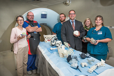
The Mayo Clinic was an early adopter of anatomical modeling and 3D printing. Jonathan M Morris, MD, a neuroradiologist and Director of Mayo’s 3D Anatomic Modeling Lab, says the hospital first used a 3D-printed model in clinical practice 14 years ago to assist in the surgical separation of conjoined twins who shared a liver. The surgeon wanted a life-size model of the organ to plan separation and reconstruction, and medical imaging played a significant role in fulfilling the request.
“Surgeons take these CT, MR, and even PET images and put them together in their mind to envision a life-sized 3D image, which I think is incredibly difficult to do and requires a lot of mental gymnastics. It’s an almost impossible task,” Dr Morris says. “3D modeling takes all that mental gymnastics work out of it and allows us to print life-sized, patient-specific anatomy and pathology from cross-sectional images.”
As the lab grew in those first few years, Dr Morris recalls, he and Jane Matsumoto, MD, a pediatric radiologist, printed between 10 and 20 3D models of various organs. Once they began printing complex oncological cases, the “barn doors opened” and there was no turning back, he says.
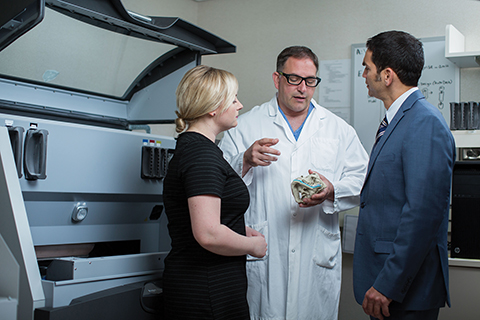
One of their most important decisions was to situate the 3D printing lab close to the surgeons, says Dr Morris, adding that the move laid the groundwork for increased collaboration and communication among the 3D printing lab team and the hospital’s surgeons, radiologists, and biomedical engineers.
“3D printing is changing the way we deliver care and solve complex problems in nearly all surgical disciplines, including orthopedic, ENT, oral and maxillofacial, neuro, thoracic, urologic, general, pediatric, and cardiac surgeries,” he says.
A New Standard for Surgical Planning
Later this year, the Mayo Clinic will open a 7,000-square-foot 3D manufacturing lab within the hospital that will boast 14 printers to meet demand. In 2019, Mayo Clinic produced 2,600 models for about 800 patients; Dr Morris projects they will print 700 custom surgical guides this year.
“3D printing of custom sterilized surgical osteotomy guides made at the point of care is now the standard of care across several surgical disciplines and pathological entities at Mayo,” Dr Morris says. “We are now doing virtual surgical planning and picking the cut planes before going to the OR and manufacturing custom plates only for a particular patient.”
In some cases, he says, surgical time has been reduced by two hours, with a savings of up to $100 per minute. Not only does 3D printing save time and money, and improve the outcomes of bony unions and oncological resections, it also allows scheduling of one additional surgical case each day.
The Mayo Clinic’s 3D Anatomic Modeling Lab includes biomedical engineers as part of the clinical team. Amy Alexander, senior biomedical engineer, and Hunter Dickin, medical 3D printing bio-medical engineer, liaise with clinicians; Dr Morris says they are critical to the success of the lab and enabling the use of 3D modeling at the point of care.
A Valuable Tool for Cardiac Surgery
Dianna M E Bardo, MD, Vice Chair of Radiology-Clinical Development at Phoenix Children’s Hospital, works closely with pediatric cardiac surgeons to treat patients with congenital heart defects. Dr Bardo says the hospital prints models for up to four cases each week.
For the last decade, she has helped moderate a 3D Experts session at the Society of Pediatric Radiology meeting. When she presents 3D virtual models on a computer, many think the next logical step would be to print a 3D model.
“Is that realistic, and do we really need all these 3D models?” she asks. “Sometimes the answer is no.”
Indeed, Dr Bardo believes 3D printing may have a greater value in producing surgical guides. For example, a ventricular septal defect is one of the hardest to close in a congenital heart defect case. A 3D surgical guide that can help the surgeon understand how the patch fits between the aorta and pulmonary aortic value, and how it separates the ventricles without making one too small, could impact how long the patient is on the bypass machine, as well as the overall surgery time, Dr Bardo says.
“While the use of 3D printing to create a prosthesis could work in orthopedic or oral and maxillofacial surgeries, it is not realistic in a moving, beating heart,” she adds. “We have had success in orthopedics by modeling a complex shoulder or hip joint where 3D printing has helped determine how to make the osteotomy.”
Patient education is another area where 3D printing can be beneficial. Dr Bardo recalls the case of a child with hypoplastic left heart syndrome. Even after multiple surgeries and imaging studies, it wasn’t until years later, when the child’s mother could hold a 3D printed model of her child’s heart in her hand, that she could fully understand the extent of her child’s condition, she says.
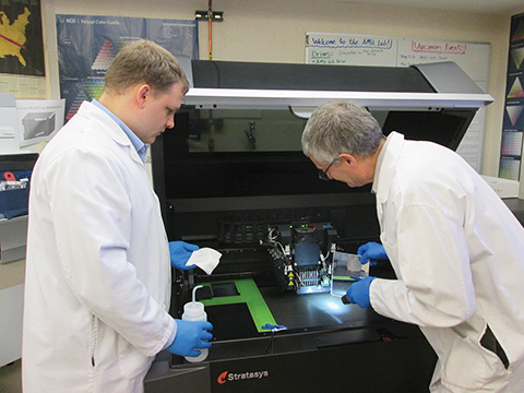
At Mallinckrodt Institute of Radiology at Washington University School of Medicine in St. Louis, meanwhile, research scientists Karthik K Tappa, PhD, and Udayabhanu M Jammalamadaka, PhD, help operate Washington University’s two 3D Printing Labs, one in the radiology department, and one in the pediatric hospital.
The lab in the radiology department actually originated at Louisiana Tech University in 2014, and was moved to Washington University in 2017 in surgical planning. Its use has evolved to include fabrication of customized, patient-specific implant and prostheses, as well as surgical guides. The estimated that 70 to 75 patient cases in 2019 involved 3D printing.
Drs Tappa and Jammalamadaka are also engaged in researching and developing novel drug delivery systems using biodegradable biopolymers. They often collaborate with Jeffery A Weisman, MD, PhD, Department of Anesthesiology, Washington University School of Medicine.
In one paper published in 2016, the two demonstrated the feasibility of using 3D printing to create a patient-tailored, bioactive hernia mesh. 2
“Some people metabolize drugs faster and using 3D printing for the delivery system we can control the dose and the shape of the implant to better deliver the drug locally, minimizing side effects and improving outcomes,” Dr Jammalamadaka explains. Their research is currently only used in animal models.
One interesting application is the case of osteomyelitis. Using 3D printing they created a drug delivery system for antibiotics that was implanted to locally attack the infection. They’ve also created 3D leads, catheters, and even an intrauterine device.
While surgical planning and surgical guides are the most common applications of 3D printing, Dr Jammalamadaka sees bioprinting as an area with significant research interest. “The final goal would be to 3D print an organ that is failing in the patient, or use 3D printing with a patient’s stem cells to address a certain condition.”
3D Printing Has Its Limitations
There are several reasons 3D printing is not more widespread and implemented mostly in major academic hospitals. For one, segmenting the region of interest (ROI) is one of the most time-consuming aspects of the process.
“Most of the available 3D software can do automatic segmentation; however, it only works when the region of interest is anatomically correct,” Dr Tappa says. “In many surgery cases, the anatomy is atypical, or not in the right shape for automation. So, we have to manually segment each layer of that image, and that takes a lot of work and hours.”
While more robust software is needed to accommodate anomalies in patient anatomy, Dr Jammalamadaka believes machine learning or deep learning could become more helpful. Even then, however, any solution needs to be adaptable to cases with atypical anatomy.
There is also the cost of the printers and consumables, says Dr Bardo. 3D printers can cost upwards of $100,000, not including the cost of consumables or the staff that must segment and print the 3D models. For reasons like these, virtual reality may prove to be a better option.
“If a surgeon wants to see a specific angle or view, or separate out anatomy, I can produce that in a virtual manner on a computer in moments and also adjust on the fly,” Dr Bardo says. “While I can cut apart a solid 3D print, it cannot be put back together and another angle or view produced without re-printing the model. I really think augmented virtual reality will become a dominant technique if it can be done without the use of heavy, disorienting goggles or cumbersome glasses. That is where I see more promise than with a 3D printed structure.”
“Augmented virtual reality allows someone who knows more about the surgical procedure to manipulate the anatomy in that virtual space,” she says. “A surgeon will want something that works with their surgical loupes, whether it is projected over the patient, in their glasses or holographically on a screen or above the patient.”
Dr Tappa agrees that the future is augmented reality. “We think 3D augmented reality will take over 3D print modeling for surgical planning,” he says. “It may be a long way to get there, but it is definitely something we see the surgeons are interested in for the future.”
Ron Schilling, PhD, has been involved in medical devices and technology for over 40 years. He currently serves as an advisor at EchoPixel, as a member of the board of directors at Histologix, as an editorial board member at Applied Radiology and an IHE (Integrating the Healthcare Enterprise) board member. Based on clinical evidence, Dr Schilling believes interactive mixed reality can bridge the interoperability gap between radiology and surgery.
“The real challenge is the interoperability across the clinical care pathway. The way to solve this is to use a common visual language to bridge that gap,” he says. “3D printing and interactive mixed reality can serve complementary roles in accomplishing this.”
Dr Schilling also sees an opportunity for interactive mixed reality to help surgeons visualize anatomy for surgical planning and also to provide 3D imaging data for a 3D printer to create the surgical guides and implants that best characterize the anatomy.
“Knowledge is the combination of cognition and intuition,” he explains. Big data, including AI and machine learning, drive cognition. The clinician adds intuition, or their perspective, to the process. Interactive mixed reality is an approach that optimizes knowledge to bridge the radiology-surgery gap.
“Interoperability across the enterprise needs to bridge that gap between specialties and clinicians, not just systems and software,” he adds. “It’s called the clinical-technical tie.”
References
- Martelli N, Serrano C, van den Brink H, et al. Advantages and disadvantages of 3Dimensional printing in surgery: A systematic review. Surgery. 2016;159(6):1485–1500. doi:10.1016/j.surg. 2015.12.017
- Ballard DH, Weisman JA, Jammalamadaka U, Tappa K, Alexander JS, Griffen FD. Three-dimensional printing of bioactive hernia meshes: In vitro proof of principle. Surgery. 2017;161(6):1479–1481. doi:10.1016/j.surg. 2016.08.033
Editor’s note: Beginning with this issue, “Radiology Matters” will replace “Technology Trends.” The goal of this new, wider-ranging department is to keep readers informed on the latest issues, trends, and technologies that are impacting medical imaging and the professionals who practice in the field.
Citation
MB M,. Radiology Matters: 3D Printing Is Bridging the Gap Between Radiology and Surgery. Appl Radiol. 2020;(2):38-41.
March 17, 2020