Photon-Counting Detector CT Shows Promise for Bone Density Data
Images

Photon-counting detector (PCD) CT spectral localizers showed clinical utility for deriving area bone mineral density (aBMD) values and, consequently, T-scores, according to a study published in the American Journal of Roentgenology (AJR).
Addressing poor adherence to dual-energy X-ray absorptiometry (DXA) screening recommendations, “the T-score derived from PCD-CT spectral localizers may serve as an opportunistic screening tool for low bone mass and osteoporosis,” noted senior and corresponding author and 2024 AJR Melvin M. Figley Fellow Francis I. Baffour, MD, from Mayo Clinic’s radiology department in Rochester, MN.
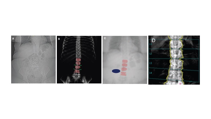
The study included patient 18 years or older scheduled for clinically indicated lumbar spine CT (October 2023-February 2024) who underwent DXA within 13 months prior or were scheduled for DXA within the subsequent 13 months. Participants underwent lumbar spine CT by PCD CT, including spectral localizer images. After extracting lumbar spine aBMD via clinical DXA reports, Baffour et al. placed ROIs on lumbar vertebral bodies and background soft tissues to extract areal densities from spectral localizer images using material decomposition. Areal densities were used to derive lumbar spine aBMD values. These aBMD values were next used to derive T-scores, classified as normal (≥-1) or abnormal (<-1) bone mass. Then, Dr. Baffour’s research team compared DXA-derived and PCD-CT measurements.
Ultimately, mean DXA-derived T-score was 0.39±1.64; mean PCD-CT derived T-score—using spectral localizer images—was 0.28±1.77 (p=.29). Lin’s concordance correlation coefficient between DXA-derived and PCD-CT T-scores was 0.90. PCD CT correctly classified 90.2% (46/51) of patients in terms of normal versus abnormal bone mass with respect to DXA.