Philips, Quibim to Collaborate on AI-enabled MR Prostate Imaging and Reporting Solution
Images
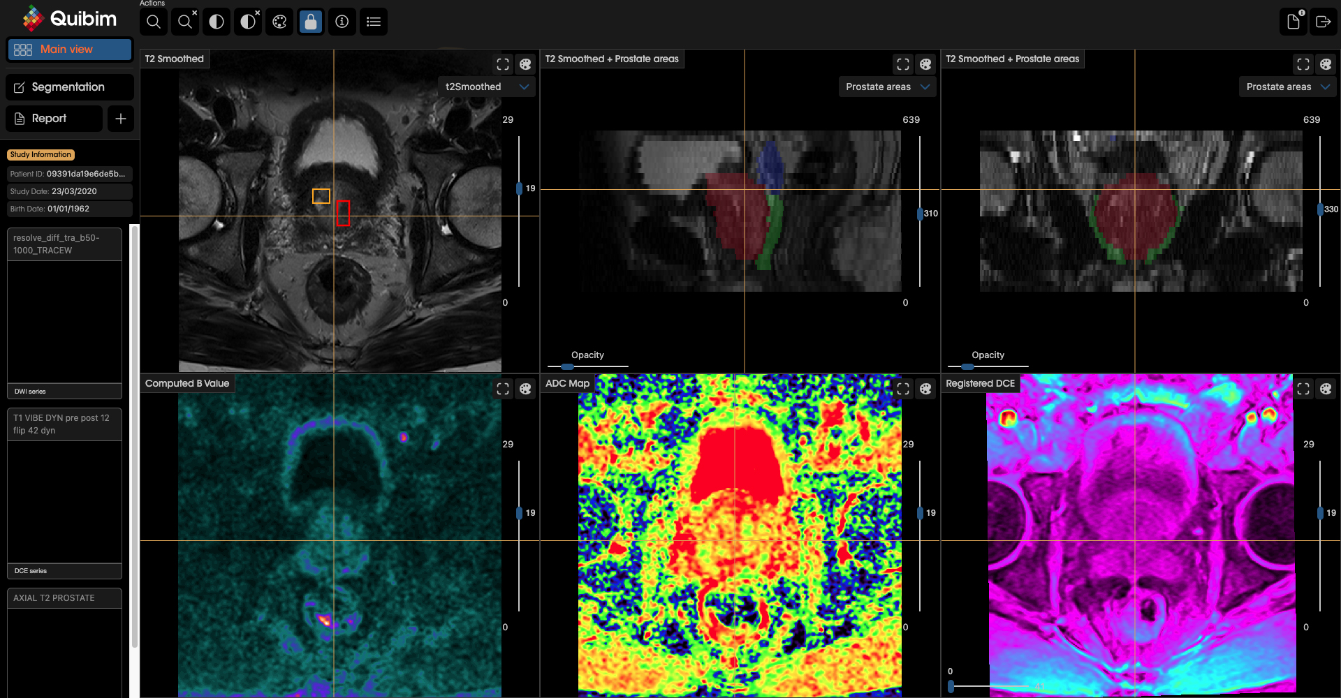
Philips and Quibim will work on an integrated solution including Quibim’s artificial intelligence (AI) based QP-Prostate software, to automate real-time prostate gland segmentation in MR images, generating meaningful quantitative insights, and standardize MR prostate exam reporting. Combined with Philips’ AI-based MR imaging techniques, the solution aims to provide clinicians with the speed and precision needed to deal with the growing number of patients in need of MR exams to help enhance diagnostic confidence, personalized treatment and better outcomes. Philips is already leveraging AI-powered technology like MR SmartSpeed image reconstruction software, which helps build clinical confidence with up to 65% higher resolution and outstanding image quality.
“This collaboration with Quibim is the latest example of our commitment to build an AI ecosystem into our Diagnostic Imaging portfolio to help detect conditions like cancer earlier, improve the rate of accurate first-time-right diagnosis, and streamline hospital operations to provide better care at lower costs,” said Ruud Zwerink, General Manager of MR at Philips. “The collaboration with Quibim will ultimately be extended to address other forms of cancer beyond prostate, where there is a need to improve efficiency and mitigate staff shortages while delivering high-quality oncology care to an increasing number of patients.”
“The collaboration including Philips’ high-speed MR imaging and Quibim’s QP-Prostate software aims to provide the speed and diagnostic confidence to support all the different steps in an integrated diagnosis, treatment, and therapy assessment workflow. The upcoming version of our lesion detection algorithm will further expand the possibilities of MRI as a gamechanger in prostate cancer screening,” said Angel Alberich-Bayarri, CEO of Quibim. “This integrated approach, combined with workflow enhancing features, is aimed at mitigating staff shortages, high burn-out rates, and cost constraints currently being experienced by many radiology and oncology departments. Patients will also greatly benefit from far less complex and painful biopsy procedures and more personalized treatment.”
“Because of its strong sensitivity to aggressive tumors, MRI has become the cornerstone of the prostate cancer diagnostic pathway. We now need to improve its specificity to avoid unnecessary biopsies, and its inter-reader reproducibility to allow reliable diagnoses outside of expert centers,” said Prof. Oliver Rouvière, Head of the Department of Radiology, Hôpital Edouard Herriot, Lyon. “Two of the biggest challenges facing any AI software dedicated to prostate MRI include the ability to demonstrate good specificity while maintaining a high level of sensitivity. Above all, we need to deliver robust diagnostic outcomes across different imaging protocols, magnetic field strengths and vendors."