Philips Announces Updates on its Development of Spectral Detector Angio CT Solution
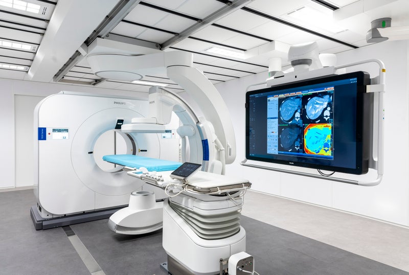 Royal Philips announced a new clinical partner and highlighted clinical studies in the development of the world’s first spectral detector angio CT solution - Philips Spectral Angio CT suite. The Spectral Angio CT combines Philips’ Spectral CT 7500 system and Image Guided Therapy System – Azurion with FlexArm – in a fully integrated interventional suite solution. With the development of this system, Philips aims to give physicians immediate access to these two key imaging modalities in a single room, enabling innovation in minimally-invasive procedures in areas such as oncology, stroke, and trauma care. The solution is currently not available in all markets or geographies, according to the company.
Leiden University Medical Center (Leiden, the Netherlands) has joined Philips’ global network of clinical partners to investigate how its spectral detector angio CT solution could potentially offer new treatment opportunities and improve patient care.
“We are excited to co-create an innovation that could play a defining role in improving patient care in the space of interventional oncology,” said Mark Burgmans, MD, Head of Interventional Radiology at Leiden University Medical Center. “Adding spectral CT imaging to the interventional suite will enable us to offer new treatment opportunities, avoid moving patients from one imaging suite to another, and offer the unique benefits of spectral CT information when you need it.”
Other leading clinical institutes that Philips is working with on this innovation are Mayo Clinic (Rochester, MN, US) and Baptist Health’s Miami Cardiac & Vascular Institute (Miami, FL, US).
At this year’s Cardiovascular and Interventional Radiological Society of Europe Annual Meeting (CIRSE 2022, September 10-14, Barcelona, Spain) a presentation will be given by Filippo Piacentino, MD, interventional radiologist at the University of Insubria (Varese, Italy), on the value of spectral CT imaging guidance for performing high-confidence tumor biopsies. The results being presented illustrate the potential for Philips’ spectral CT technology to better guide biopsies by distinguishing between active and non-active regions in a tumor. Ensuring that a biopsy contains a high number of actively dividing cancer cells is important for high-confidence diagnosis.
“With conventional CT, large masses may appear as a largely uniform mass, making highly targeted biopsy difficult,” said Dr Piacentino. “By fusing images from Philips’ XperGuide live needle guidance with images from spectral CT, that are color-coded based on the effective atomic number of tissues and provide a large amount of additional information, we can now investigate the possibility of obtaining better defined biopsy targets with a fewer number of inconclusive biopsies.”
Philips also today announced a research collaboration with the University of Pennsylvania (Pennsylvania, PA, US) to study the practicality of using spectral CT-based tissue temperature mapping to provide real-time feedback during tumor thermal ablation procedures to confirm its effectiveness before the patient leaves the room. This will potentially reduce the risk of localized tumor recurrence.
“Announcing these important milestones in the development of our unique Philips Spectral Angio CT suite shows our strong commitment and progress in co-creating the future of image-guided therapy,” said Karim Boussebaa, General Manager of Image Guided Therapy Systems at Philips. “By combining the best of our award-winning modalities into a single suite we aim to unlock new treatment approaches that could benefit both patients and their physicians.”