PET/MR Recommended for Diagnosing Chronic Osteomyelitis, if Available
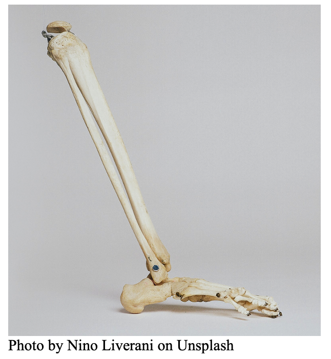 A study published in the European Journal of Hybrid Imaging recommends the use of PET/MR, if available, over PET/CT for diagnosing osteomyelitis.
The retrospective analysis of 36 patients with 18F-FDG PET/MRI scans for suspected osteomyelitis was performed by both a nuclear medicine physician and a radiologist. Sensitivity, specificity, and accuracy were determined with the clinical assessment by an orthopedic surgeon, based on subsequent intraoperative microbiology or long-term follow-up, as the ground truth. Standardized uptake values (SUV) were measured and analyzed by means of receiver operating characteristics (ROC).
The sensitivity, specificity, and accuracy of 18F-FDG PET/MRI was 78%, 100%, and 86% respectively while the area under the ROC curve was .736, .755, and .769 for the SUVmax, target to background ratio, and SUVmax_ratio respectively. The authors reported these values were in a similar range and not statistically different compared to diagnostic value for 18F-FDG PET/CT imaging of osteomyelitis as found in the literature.
Imaging plays an important role in the diagnostic work-up of osteomyelitis. The authors noted that their study found specificity to be higher than sensitivity while other studies found sensitivity higher than specificity. They suggest, “the accuracy of an imaging modality might be the most important parameter for the clinician planning the treatment strategy of chronic osteomyelitis because of its chronic nature. Our accuracy of 86% is among the highest of the reported studies (range 67–91%).”
With the added benefit of a reduced radiation dose and additional soft tissue information for surgical treatment planning when using PET/MR versus PET/CT, the authors advise physicians with PET/MR to consider using it rather than PET/CT in osteomyelitis diagnosis.