No MRI-Detectable Brain Injury Found in People With “Havana Syndrome”
Images
A research team at the National Institutes of Health (NIH) found no significant evidence of MRI-detectable brain injury, nor differences in most clinical measures compared to controls, among a group of federal employees who experienced anomalous health incidents (AHIs). These incidents, including hearing noise and experiencing head pressure followed by headache, dizziness, cognitive dysfunction and other symptoms, have been described in the news media as “Havana Syndrome” since US government personnel stationed in Havana first reported the incidents. Scientists at the NIH Clinical Center conducted the research over the course of nearly five years and published their findings in two papers in JAMA.
“Our goal was to conduct thorough, objective and reproducible evaluations to see if we could identify structural brain or biological differences in people who reported AHIs,” said Leighton Chan, MD, chief, rehabilitation medicine and acting chief scientific officer, NIH Clinical Center, and lead author on one of the papers. “While we did not identify significant differences in participants with AHIs, it’s important to acknowledge that these symptoms are very real, cause significant disruption in the lives of those affected and can be quite prolonged, disabling and difficult to treat.”
Researchers designed multiple methods to evaluate more than 80 US government employees and their adult family members, mostly stationed abroad, who had reported an AHI and compared them to matched healthy controls. The control groups included healthy volunteers who had similar work assignments but did not report AHIs. In this study, participants underwent a battery of clinical, auditory, balance, visual, neuropsychological and blood biomarkers testing. In addition, they received different types of MRI scans aimed at investigating volume, structure and function of the brain.
In this study, researchers obtained multiple measurements and used several methods and models to analyze the data. This was done to ensure the findings were highly reproducible, meaning similar results were found regardless of how many times participants were evaluated or their data statistically analyzed. Scientists also used deep phenotyping, which is an analysis of observable traits or biochemical characteristics of an individual, to assess any correlations between clinically reported symptoms and neuroimaging findings.
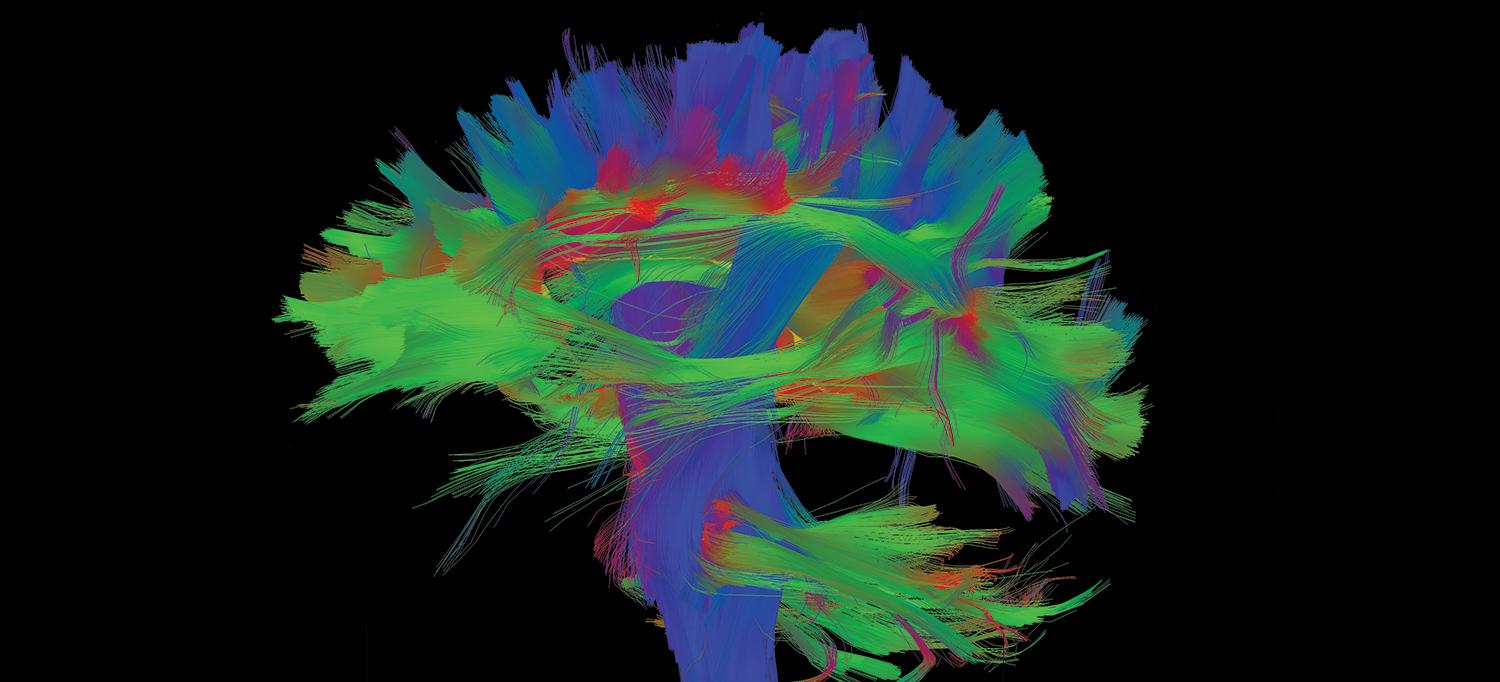
For the imaging portion of the study, participants underwent MRI scans an average of 80 days following symptom onset, although some participants had an MRI as soon as 14 days after reporting an AHI. Using thorough and robust methodology, which resulted in highly reproducible MRI metrics, the researchers were unable to identify a consistent set of imaging abnormalities that might differentiate participants with AHIs from controls.
“A lack of evidence for an MRI-detectable difference between individuals with AHIs and controls does not exclude that an adverse event impacting the brain occurred at the time of the AHI,” said Carlo Pierpaoli, MD, PhD, senior investigator and chief of the Laboratory on Quantitative Medical Imaging at the National Institute of Biomedical Imaging and Bioengineering, part of NIH, and lead author on the neuroimaging paper. “It is possible that individuals with an AHI may be experiencing the results of an event that led to their symptoms, but the injury did not produce the long-term neuroimaging changes that are typically observed after severe trauma or stroke. We hope these results will alleviate concerns about AHI being associated with severe neurodegenerative changes in the brain.”
Similarly, there were no significant differences between individuals reporting AHIs and matched controls with respect to most clinical, research and biomarker measures, except for certain self-reported measures. Compared to controls, participants with AHIs self-reported significantly increased symptoms of fatigue, post-traumatic stress and depression. Forty-one percent of participants in the AHI group, from nearly every geographic area, met the criteria for functional neurological disorders (FNDs), a group of common neurological movement disorders caused by an abnormality in how the brain functions, or had significant somatic symptoms. FNDs can be associated with depression and anxiety, and high stress. Most of the AHI group with FND met specific criteria to enable the diagnosis of persistent postural-perceptual dizziness, also known as PPPD. Symptoms of PPPD include dizziness, non-spinning vertigo and fluctuating unsteadiness provoked by environmental or social stimuli that cannot be explained by some other neurologic disorder.
“The post-traumatic stress and mood symptoms reported are not surprising given the ongoing concerns of many of the participants,” said Louis French, PsyD, neuropsychologist and deputy director of the National Intrepid Center of Excellence at Walter Reed National Military Medical Center and a co-investigator on the study. “Often these individuals have had significant disruption to their lives and continue to have concerns about their health and their future. This level of stress can have significant negative impacts on the recovery process.”
The researchers note that if the symptoms were caused by some external phenomenon, they are without persistent or detectable patho-physiologic changes. Additionally, it is possible that the physiologic markers of an external phenomenon are no longer detectable or cannot be identified with the current methodologies and sample size.
Related Articles
Citation
No MRI-Detectable Brain Injury Found in People With “Havana Syndrome”. Appl Radiol.
March 23, 2024