New Soft Tissue on CT Suggests Local Recurrence After Pancreatic Cancer Resection
Images

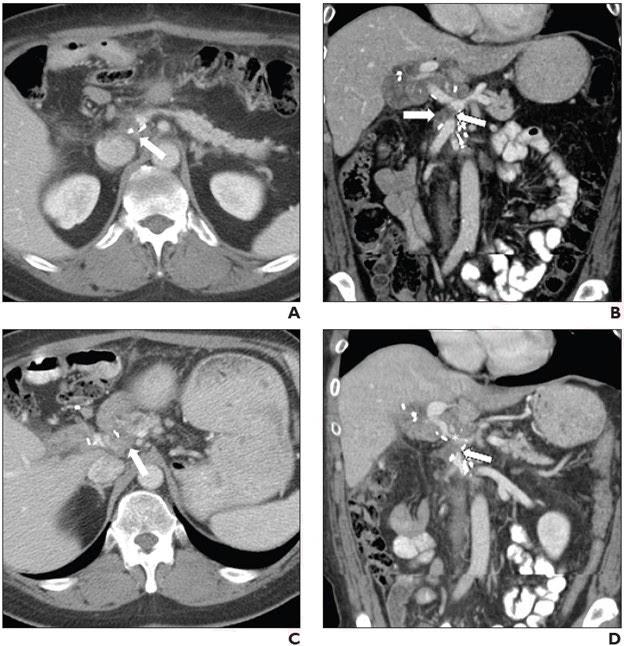
A study published in the American Journal of Roentgenology (AJR) highlights the role of soft tissue, particularly when associated with vessel encasement and luminal narrowing, in raising suspicion for local recurrence (LR) after pancreatic ductal adenocarcinoma (PDAC) PDAC resection.
“This AJR study provides insight into findings described in the Society of Abdominal Radiology’s (SAR) PDAC Disease-Focused Panel (DFP) consensus statement that indicate the presence, or likely development, of postresection LR, while highlighting opportunities for continued optimization,” wrote corresponding author Tae-Hyung Kim, MD, MS, from the radiology department at Memorial Sloan Kettering Cancer Center in New York City.
Kim et al.’s study included 126 patients (mean age, 68.5 years; 72 men, 54 women) who underwent Whipple surgery for PDAC (January 2009–December 2014). Three radiologists independently reviewed baseline and subsequent postoperative contrast-enhanced abdominopelvic CT examinations performed within 2 years postoperatively, evaluating features in the SAR PDAC DFP consensus statement relating to surgical bed stranding, surgical bed soft tissue, vessel encasement, main pancreatic duct dilatation, and ascites. After calculating interreader agreement, the reference standard for LR development within 2 years postoperatively incorporated all available information. Imaging features’ frequencies were calculated for recurrence examinations (i.e., first surveillance examinations indicating LR).
Ultimately, on the first surveillance CT examinations showing LR after PDAC resection, across three readers, new or increased soft tissue was present in 80–86%, and soft tissue with vessel encasement and luminal narrowing in 36–59%. These findings showed moderate and fair interreader agreement, respectively.