New Images Show CT Cinematic Rendering of SARS-CoV-2 Pneumonia
New images published in Radiology show PCR-positive SARS-Cov-2 pneumonia using cinematic rendering of CT images, a new way to present the three dimensionality of the various densities contained in volumetric CT data.
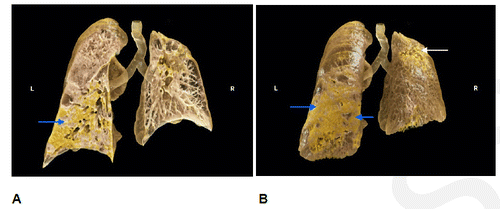
Figure A shows cinematic rendering of a chest CT from a 60-year-old male presenting with RT-PCR positive SARS-CoV-2 pneumonia showing a coronal cross-section with consolidation in the left lower lobe (arrow). Figure B shows coronal shaded surface display after cinematic rendering of the same patient.
This case is from the RSNA-RICORD dataset1,2 that shows the typical presentation of SARS-Cov-2 pneumonia with ground-glass subpleural opacities that are clearly seen. The higher attenuation of lung tissue filled with fluid results in these areas appearing patchy or spongy.
A video animation sequence of coronal shaded surface display after cinematic rendering also shows a coronal cross-section through both lungs and 3D rotation around each lung separately.
Cinematic Rendering is a digital 3D visualization technique that converts grayscale slices from CT or MRI into colored 3D volumes via transfer functions illuminating the reconstruction with physical light simulation. This produces a natural, photorealistic image that is intuitively understandable and can be used for clinical purposes.
The SARS-Cov-2 pandemic has spread rapidly throughout the world since its first reported infection in Wuhan, China. Despite the introduction of vaccines for this important viral infection, there remains a significant public health risk to the population as this virus continues to mutate. While it remains unknown if these new mutations will evade the current vaccines, it is possible that we may be living with this infection for many years to come as it becomes endemic.
Software development and access provided in cooperation with Siemens Healthineers GmbH, Erlangen.
See full images and video at https://pubs.rsna.org/doi/10.1148/radiol.212902.
1. Tsai EB, Simpson S, Lungren MP, Hershman M, Roshkovan L, Colak E, Erickson BJ, Shih G, Stein A, Kalpathy-Cramer J, Shen J, Hafez M, John S, Rajiah P, Pogatchnik BP, Mongan J, Altinmakas E, Ranschaert ER, Kitamura FC, Topff L, Moy L, Kanne JP, Wu CC. The RSNA International COVID-19 Open Radiology Database (RICORD). Radiology2021;299(1):E204–E213. doi: https://doi.org/10.1148/radiol.2021203957Link, Google Scholar
2. Clark K, Vendt B, Smith K, Freymann J, Kirby J, Koppel P, Moore S, Phillips S, Maffitt D, Pringle M, Tarbox L, Prior F. The Cancer Imaging Archive (TCIA): maintaining and operating a public information repository. J Digit Imaging2013;26(6):1045–1057. doi: https://doi.org/10.1007/s10278-013-9622-7Crossref, Medline, Google Scholar
Citation
New Images Show CT Cinematic Rendering of SARS-CoV-2 Pneumonia. Appl Radiol.
December 22, 2021