Monitoring Cerebral Blood Flow With Ultrasound in Newborns Undergoing Surgery Prevents Brain Damage
Images
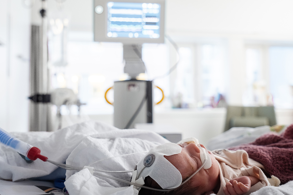
Researchers and neonatal specialists at Norwegian University of Science and Technology (NTNU) and St. Olavs Hospital are behind the first study in which ultrasound is used to monitor cerebral blood flow in newborn babies under general anesthesia. Ultrasound technology from NTNU makes it possible to monitor cerebral blood flow in newborn babies, helping prevent brain damage in premature and sick infants who require surgery.
“We have always believed that this could revolutionize neonatal care. Everything indicates that we are closer to being able to prevent brain damage in premature infants and sick newborns,” says pediatrician Sigrid Dannheim Vik, who is doing a PhD on NeoDoppler technology.
Over a two-year period, she has used the equipment to measure cerebral blood flow in 30 newborn babies under general anesthesia. Some of them were born very prematurely. Others came into the world around their due date, but were born with congenital defects that had developed in the womb.
Most of the patients had gastrointestinal complications, such as lack of passage through the small intestine. Premature babies often have an immature intestine that is easily perforated in the neonatal period.
“These babies require surgery – often acutely. There is no other choice,” says Vik.
The longest courses of continuous ultrasound measurements lasted 10-11 hours, and some of her patients weighed less than 1000 grams.
Babies who are born prematurely are prone to brain damage in the neonatal period and are very vulnerable to blood pressure fluctuations. The risk increases if they have to undergo surgery.
The aim of the NTNU study has been to use the new ultrasound technology to see how cerebral blood flow changes while the infants are under general anesthesia.
The conclusion is that NeoDoppler provides doctors with access to critical information that has previously been unavailable.
“The equipment provides important additional information compared with today’s standard monitoring equipment,” Vik said.
Currently, indirect factors such as blood pressure, pulse measurements and general clinical assessments are used to see if the child has adequate blood circulation.
“We don’t actually have a reliable measurement method for monitoring the most important organ, namely the brain,” says the pediatrician and researcher.
NeoDoppler measurements are taken continuously before, during and after surgery. The equipment is able to instantly detect changes in cerebral blood flow. As a result, measures that can prevent brain damage and that are adapted to the individual child can be implemented immediately.
It also helps doctors make individual assessments and tailor treatment, rather than having to rely on general recommendations.
“I am in no doubt that we are now moving closer to preventing brain damage.”
The researchers believe NeoDoppler can provide a new standard in monitoring newborn babies.
“The problem today is that we don’t know what the most favorable blood pressure in newborns is with regard to maintaining stable blood circulation in the brain, and it probably varies from one infant to the next,” says Sigrid Dannheim Vik.
What she and her colleagues saw, and which they believe gives cause for concern, was that cerebral blood flow speed was more than halved during anesthesia compared with when the patients were awake.
“Our findings indicate that newborns who are placed under general anesthesia should probably have a higher blood pressure than is currently recommended. The current reference range should probably be higher,” says Vik.
The NeoDoppler ultrasound probe is the shape of a small button and has a diameter of around 1 centimeter. It can be attached to a cap and placed gently over the fontanelle (the soft spot at the top of a baby’s head).
The fontanelle closes towards the end of the first year of life when the bones of the skull fuse together. Ultrasound is not able to pass through bone. Therefore, the fontanelle is like a window that is only open for a limited period of time, through which doctors can directly monitor blood flow.
More clinical trials are needed before NeoDoppler becomes standard monitoring equipment in neonatal care. These will be accurate with regard to effect, but the children must be followed up over a longer period of time in order to show that it helps prevent brain damage.
Paediatric cardiologist and researcher Siri Ann Nyrnes is behind the invention, together with Professor Hans Torp. She is also Sigrid Dannheim Vik’s main academic supervisor.
“The NeoDoppler system is CE certified and can therefore be used in multiple environments. Nyrnes says that the ultrasound is now being tested by partners at Oslo University Hospital, UMC Utrecht in the Netherlands and at the Hospital for Sick Children in Toronto, Canada.