Method to Trigger and Image Seizures May Help Guide Epilepsy Seizures
Images
Researchers have developed a new method for triggering and imaging seizures in epilepsy patients, offering physicians the ability to collect real-time data to tailor epilepsy surgery, according to research published in The Journal of Nuclear Medicine. In contrast to previous practice, where physicians from neurology and nuclear medicine had to wait for hours to days in hopes of capturing the onset of a seizure, the new method is convenient, spares resources, and is clinically feasible.
People with epilepsy and seizures who do not respond to medication are often helped by brain surgery. The goal of the surgical procedure is to remove the epileptic brain tissue and spare the healthy brain tissue to control seizures but avoid neurological deficits. “Precisely delineating the epileptic brain tissue is essential for successful surgeries, and obtaining timely images of seizures may help formulate surgical plans with increased precision” said Sabry L. Barlatey, MD, PhD, resident in the Department of Neurosurgery at University Hospital of Bern in Bern, Switzerland.
The ictal SPECT method has been used since the 1990s as the sole neuro-imaging technique able to capture an image of an epileptic seizure propagating in the brain. However, due to the growing cost and time constraints in health care, most epilepsy centers abandoned this potentially informative technique.
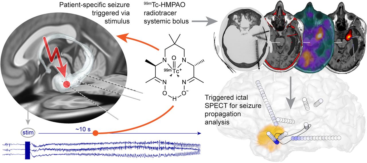
“In this study, instead of waiting for spontaneous occurrences, we imaged planned seizures that were triggered with targeted electrical stimulation to the brain. To our knowledge, this simple idea had never been tested before,” noted Maxime O. Baud, MD, PhD, professor of neurology in the Department of Neurology at the University Hospital of Bern.
Three adult participants with left temporal epilepsy were included in the case study. Authors identified and used stereotactic electroencephalography (sEEG) leads in targeted cerebral areas to trigger patient-typical seizures. The radiotracer 99mTc-HMPAO was administered within 12 seconds of ictal onset and SPECT images were acquired within 40 minutes.
Seizures were successfully triggered in each participant, replicating the patient-typical seizure semiology and electrographic pattern on sEEG without any adverse events. Each triggered seizure was patient-specific, and the imaged early seizure propagation was unique. In the first two cases, ictal SPECT offered complementary information to sEEG and revealed early involvement of brain areas lacking electrode coverage. In the third case, sEEG and ictal SPECT provided overlapping information.
“The finding of this study is of practical nature, as it greatly facilitates the acquisition of the ictal SPECT,” noted Thomas Pyka, MD, Privatdozent in the Department of Nuclear Medicine at the University Hospital of Bern. “This may help obtain images of greater quality and could contribute to the refinement of resection planning, improving seizure and cognitive outcomes in epilepsy surgery.”