Metal Implants and Vertical-Field MRI: To Scan or Not to Scan?
 VIENNA, AUSTRIA – With its powerful magnetic fields, MRI has always presented challenges when it comes to scanning patients with metallic implants, and these challenges are particularly acute for users of vertical-field MR systems. Fortunately, consensus is evolving that these scanners are suitable for patients with passive metallic implants that have been labeled at 1.5- and/or 3-Tesla.
That’s according to a March 1 symposium by Frank Shellock, PhD, at the European Congress of Radiology (ECR) on the benefits of vertical field MR systems. Dr. Shellock is a professor and the director of MRI Safety at the University of Southern California and runs the website MRISafety.com.
While high-field, horizontal-bore superconducting systems get most of the attention in radiology, vertical-field scanners have many advantages. For example, these scanners have a more open architecture – a major benefit when scanning claustrophobic or anxious patients.
But one drawback of vertical-field MRI is that most of the implant testing has been almost exclusively performed on horizontal-bore, high-field MRI systems, says Dr Shellock. MRI facilities already conduct extensive screening of patients prior to MRI scans to ensure they can be examined safely. When it comes to implants, several resources are available for MRI users to review – such as MRISafety.com – to determine their suitability for patients referred for MRI exams.
These tests focus on four major areas that can cause potential complications during MRI exams: force, torque, heating, and artifacts. In the US, results of implant testing are forwarded to the US Food and Drug Administration (FDA), which has three labeling categories for implants: MR Safe, MR Conditional (meaning patients can be scanned under certain conditions to ensure safety), and MR Unsafe.
But can the results of implant testing conducted on high-field, horizontal-bore MR scanners be automatically translated to vertical-field systems? Such scanners behave differently from supercons, notes Dr Shellock. For instance, the direction of the static magnetic field is different, and some issues common in superconducting MRI – such as MRI-related heating of implants – are not as much of a problem on vertical-field MR systems. Additionally, vertical-field MR systems operating below 1.5 Tesla inherently generate fewer susceptibility artifacts around metallic implants compared with horizontal-field scanners.
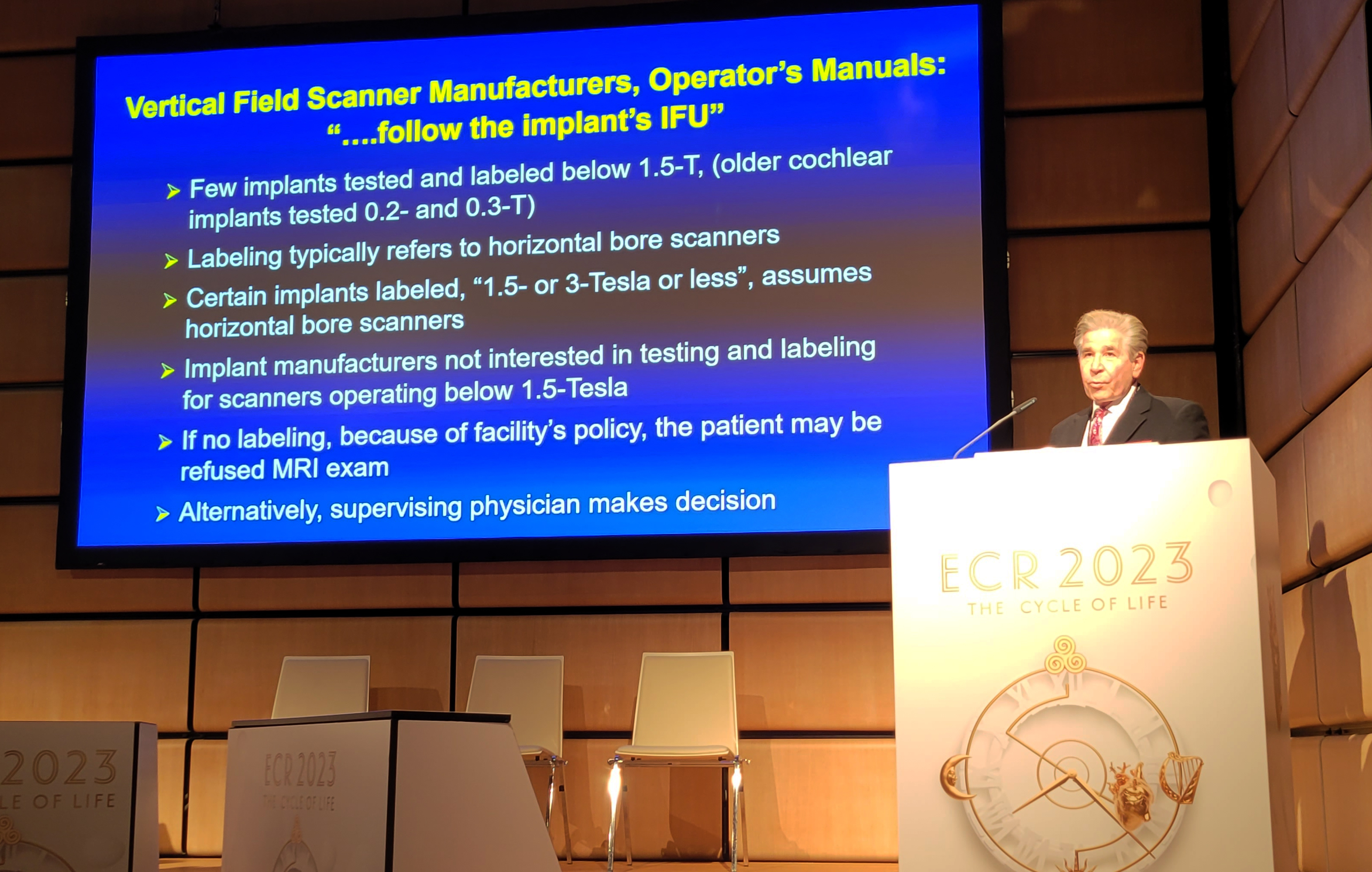
The labeling of an implant isn’t much help either, Dr Shellock says. Most implants are tested and labeled at 1.5- and/or 3-Tesla, and implant manufacturers aren’t interested in testing their devices at lower field strengths (although this may be beginning to change, he notes). As a result, many MRI facilities are adopting general policies indicating that it’s acceptable to scan a patient who has a passive metallic implant in a vertical-bore MR system, if the implant has been labeled at 1.5- and/or 3-Tesla, thus, ensuring that patients are not turned away unnecessarily.
“With that general policy in place, you can effectively scan patients with the thousands of implants that have been tested and labeled at 1.5- and/or 3-Tesla,” he says. “That would allow you to effectively utilize all of the proposed advantages of vertical-bore systems operating from 1.2 down to 0.2 Tesla.”
But what about patients who have untested passive implants, such as orthopedic implants? Some of these devices, such as hip prostheses, are among the largest implants in patients. For these individuals, the best choice is for the supervising physician to decide case by case, he suggests, with consideration given to the field strength and the scanner’s transmit frequency.
“Keep in mind that the direction of the E-fields will result in lower temperature rises,” Dr Shellock says. “You can also then afford a larger margin of safety if there is any possible concern for MRI-related heating of an unlabeled, untested passive implant.”
In addition, implant manufacturers may be paying more attention to labeling implants specifically for vertical-field magnets, he notes. For instance, Medtronic has included text for one of its cardiac monitoring devices indicating that it is acceptable for scanning in a vertical-bore system.
Several other presentations in the ECR symposium discussed other advantages of vertical-bore scanners. In one talk, Mihaly Aradi, PhD, from Hungary discussed his facility’s experiences with a 0.4-Tesla vertical field scanner. The system has a number of advantages in a country like Hungary, such as a lower operating cost – the scanner’s electricity usage is one-tenth that of a superconducting magnet, he notes. It also has advantages due to its open design, which makes it more suited to patients with claustrophobia. Dr Aradi’s facility has been operating the scanner for years and continues to use it, even after acquiring a state-of-the-art 3-Tesla scanner.
In another talk, Dr Juergen Biederer of Germany discussed benefits of vertical-bore scanner technology, in this case a 1.2-Tesla scanner with an open design. Such systems meet patient desires for a scanning environment that’s not claustrophobic and offer more flexibility in patient positioning. He cited a study conducted in Germany on the economic advantages of such a system, noting that a scan aborted due to patient claustrophobia can cost an imaging facility nearly 6,000 euros in lost opportunity costs.
Citation
Metal Implants and Vertical-Field MRI: To Scan or Not to Scan?. Appl Radiol.
March 2, 2023