Imaging With FAPI PET Accurately Assesses Rheumatoid Arthritis Disease Activity
Images

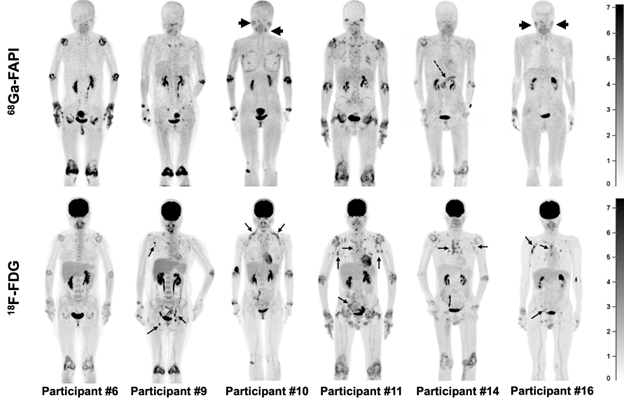
New research presented at the 2023 Society of Nuclear Medicine and Molecular Imaging Annual Meeting shows that PET imaging with 68Ga-FAPI offers a feasible, reliable, and reproducible method for evaluating disease activity in rheumatoid arthritis. 68Ga-FAPI PET/CT imaging of joint involvement correlated with clinical and laboratory assessments of disease activity. It also showed a greater number and degree of affected joints when compared to imaging with 18F-FDG PET/CT.
In rheumatoid arthritis, management strategies aim to control inflammation and prevent joint injury. Frequent assessment of disease activity, as often every three months, is essential to maintaining control of the disease. Clinical assessment of disease activity usually includes physical examination of the joints, acute-phase reactant levels, and patient reporting of disease activity and quality of life. However, physical examination and patient self-reporting may lack reproducibility in clinical practice.
“We know that fibroblast activation protein (FAP) is overexpressed in rheumatoid arthritis,” said Yaping Luo, MD, assistant professor at Peking Union Medical College Hospital, Beijing, China. “Our team sought to leverage this FAP overexpression with PET imaging to improve assessment of disease activity in rheumatoid arthritis patients.”
The study was designed to determine the performance of 68Ga-FAPI in assessing joint disease activity in rheumatoid arthritis patients and to compare it with 18F-FDG imaging. Twenty rheumatoid arthritis patients with moderate to high disease activity underwent clinical and laboratory assessments as well as dual-tracer PET/CT imaging with 68Ga-FAPI and 18F-FDG. Images derived from the PET/CT imaging were analyzed for correlation to clinical and laboratory assessments of disease activity. The two imaging methods were also compared.
When combining physical examination and PET/CT, a total of 314 joints showed disease activity. Among them, 167 joints were positive on both physical examination and 68Ga-FAPI PET/CT. The positive rate of detecting involved joints was 75.5% for physical examination and 77.7% for 68Ga-FAPI PET/CT. The matching rate of physical examination and 68Ga-FAPI PET was 53.2%. 68Ga-FAPI PET was also positively correlated with other clinical and laboratory assessments.
The output of both PET/CT imaging techniques (68Ga-FAPI and 18F-FDG) detected 244 affected joints, all of which were positive on 68Ga-FAPI; 15 of these joints were not detected by 18F-FDG. In addition, the maximum standardized uptake value of the most affected joint in each participant was significantly higher with 68Ga-FAPI PET than with 18F-FDG PET.
“This study offers a reliable, reproducible imaging technique that can be used in the evaluation of disease activity in rheumatoid arthritis,” said Luo. “In addition to detecting disease activity, the depiction of 68Ga-FAPI in patients with rheumatoid arthritis may help researchers better understand the role of fibroblast-like synoviocyte cells that are involved in the inflammation of the cartilage and bone in rheumatoid arthritis.”