FDA Clears MAGNETOM Terra.X 7T MR Scanner
Images
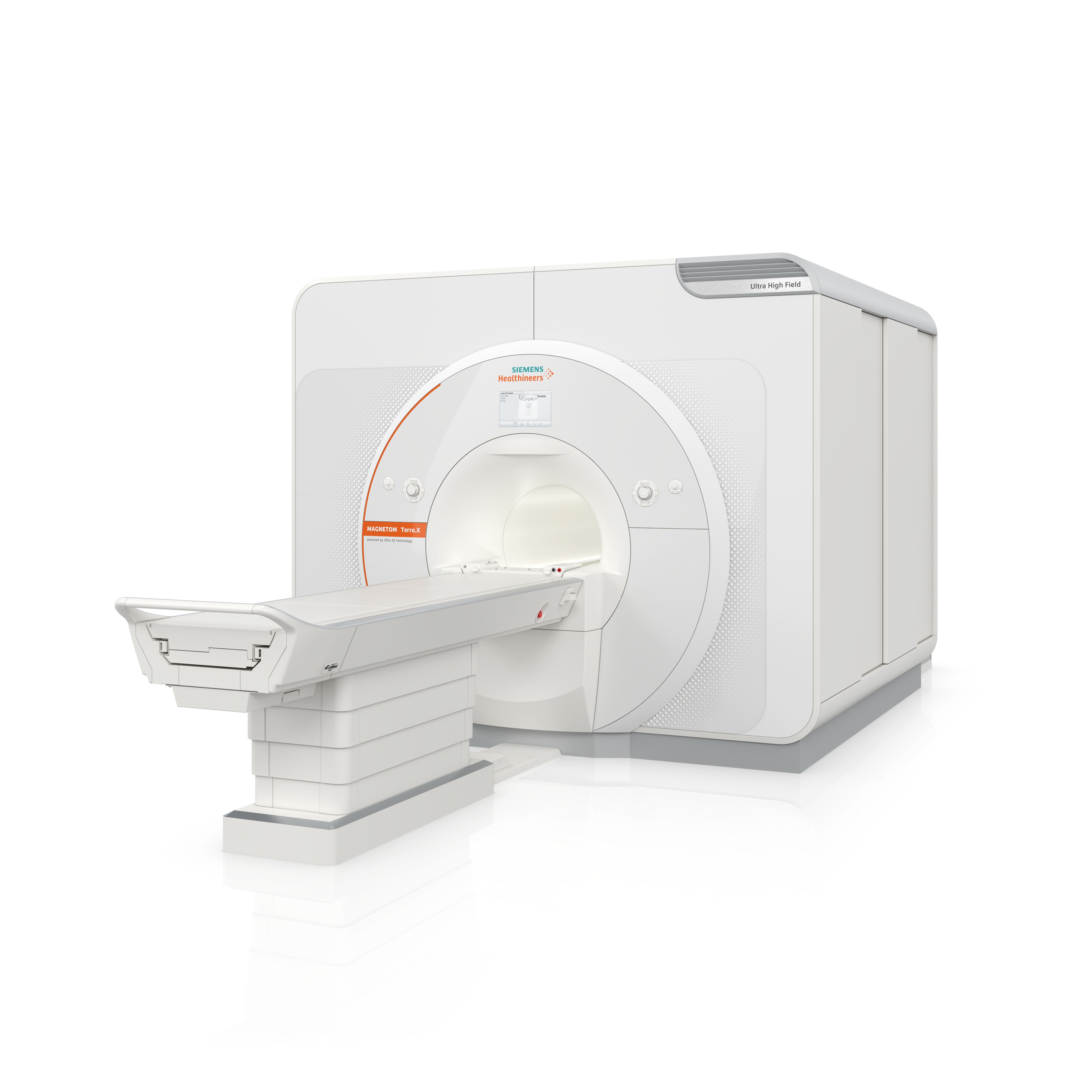
Siemens Healthineers announced US FDA clearance of the MAGNETOM Terra.X, a second-generation clinical 7T MR scanner featuring new hardware and XA60A software. The MAGNETOM Terra.X is the successor to the MAGNETOM Terra, which debuted in 2017 as the world’s first 7T clinical MR scanner, enabling extremely high-resolution imaging. An upgrade to the MAGNETOM Terra.X for installed MAGNETOM Terra systems was included in the FDA clearance, as was an upgrade for the MAGNETOM Terra to the XA60A software version.
“The MAGNETOM Terra.X builds on our engineering achievements as pioneers in 7T MRI by providing the ultra-high-field community with the necessary tools to offer higher-quality patient care not just in neurological imaging, but also in the knee,” said Katie Grant, vice president of Magnetic Resonance at Siemens Healthineers North America. “Additionally, this scanner supports key research initiatives pertaining to neurodegenerative disease, which is a major concern for our aging population.”
The new Ultra IQ Technology of the MAGNETOM Terra.X introduces an unprecedented level of image quality for 7T scanning. The world’s first clinical eight-channel parallel transmit architecture enables a degree of image clarity that reveals even extremely subtle pathological details.
The MAGNETOM Terra and MAGNETOM Terra.X with syngo MR XA60A software have Deep Resolve, the company’s deep learning-based image reconstruction technology that has been trained specifically with 7T data to deliver new levels of speed and image resolution at 7T. For example, the combination of Deep Resolve and accelerated acquisition techniques such as high parallel acquisition can enable a routine high-resolution brain and knee exam in under 20 minutes, which is a dramatic improvement for 7T. Additionally, the scanner’s new, high-performance gradient system delivers exceptional diffusion imaging and functional MR imaging.
The scanner’s multinuclear option permits clinical imaging of substances in the body—for example, sodium in the brain and phosphorous in muscle tissue—that are not as easily visualized with standard MR field strengths. The ability to perform this type of imaging adds a metabolic component to the already exceptional anatomical imaging capabilities of 7T MR.