Enhancing Diagnostics and Patient Care with Spectral CT
Images


BROUGHT TO YOU BY PHILLIPS.
Few radiologists could imagine current clinical practice without CT. This powerful diagnostic tool’s ease of use and ubiquitous availability only fortify its significance. Nevertheless, traditional CT often still yields inconclusive findings that require supplemental testing to achieve a confident diagnosis.
Philips IQon Spectral CT was created to address that challenge. Since its introduction, the IQon Spectral CT has had a profound effect on clinicians’ ability to detect and diagnose disease with a single exam, stratifying high energy and low energy photons simultaneously and using color to characterize the material content of critical structures. The ability to collimate by energy has elevated CT scanning beyond “tomodensity” to the new, yet predicted by Hounsfield himself, field of “tomochemistry.” By harnessing the advances in this technology, clinicians have taken CT to a whole new level of enabling diagnoses and, in some cases, making diagnoses faster.
Contrast dose matters
The Philips IQon Spectral CT provides a more complete picture of the patient with its ability to visualize cardiac abnormalities and lung capacity, which can be crucial to planning successful surgeries, such as transcatheter aortic valve replacement. Additionally, it permits a more comprehensive risk assessment using lower amounts of iodinated contrast.
The patients receiving these types of treatments are typically older, often have comorbidities such as renal dysfunction, and are not amenable to routine open surgical procedures like an open sternotomy or aortic valve replacement, said Amit Gupta, MD, Cardiothoracic Radiologist at University Hospitals, Cleveland Medical Center, and Assistant Clinical Professor of Radiology, Case Western Reserve University School of Medicine.
“For our high-risk patients needing valve replacement who may be poor candidates for open surgical procedures,” explained Dr. Gupta, “we can treat them by going through the femoral artery and placing the aortic valve endovascularly, rather than opening up that patient. That’s why reducing the contrast dose is so important.
“Using the virtual monoenergetic images on the IQon Spectral CT, we are able to substantially reduce the contrast dose for patients who may already be struggling with renal function, giving these patients less exposure and possibly fewer complications,” he added. “So rather than doing a CT angiogram, chest, abdomen and pelvis with more than 100mL of contrast, we do it with only 50mL.”
Instances of contrast dose extravasation also occur, though infrequently, and these patients will frequently need to be re-imaged, resulting in even more radiation exposure and a second dose of contrast. Chip Truwit, MD, Chief of Radiology at Hennepin County Medical Center (Hennepin), explained that he utilizes Philips IQon Mono-E spectral reconstruction in these cases.
“Using IQon Spectral CT technology, I never have to repeat that exam,” Dr. Truwit said. “Typically, as many as 10% of our patients may experience extravasation due to fragile veins, and this is not unique to our facility. If we were using conventional CT, we would probably have to repeat the exam. With IQon Spectral CT, you just need to adjust the image to 40 keV and essentially, the study is salvageable. It saves time, cost, and patient dose, making it much better all around.”
Sustainable changes
Dr. Gupta worked exclusively on clinical research before his facility received its clinical IQon Spectral CT in April 2016. Since then, he has replaced traditional CT with the IQon in a number of different protocols because of the benefits it provides.
“We’ve put our IQon Spectral CT to use beyond the mere evaluation of coronary stenosis and utilize it for more accurate plaque characterization and assessment of myocardial perfusion,” Dr. Gupta said. “We’ve actually changed a lot of our protocols since we began using the IQon. All of our patients undergoing transcatheter aortic valve implantation (TAVI) will have their planning exam done with IQon Spectral CT, as well as those patients having WATCHMAN device placement for atrial fibrillation. These are always done on our spectral CT now.”
Hennepin was one of the first facilities to experience IQon Spectral CT when it was introduced, uniquely placing the unit in the Emergency Department (ED) and using its advanced diagnostic capabilities to help clinicians stabilize patients. When the opportunity arose, a second IQon Spectal CT was sited in Hennepin’s new ambulatory facility.
“We’re using the IQon Spectral CT now on every chest, abdomen, and pelvis case we can,” explained Dr. Truwit. “It’s definitely transformed our ED practice. CT is often the difference between life and death, between getting to the diagnosis and not, when time is of the essence. And with the IQon we don’t have to do anything differently because it’s the detector that’s different. You get the spectral data on every patient and you decide when you need it. Now we’re using IQon Spectral CT in our outpatient facility; we now scan over 70% of our CT patients using spectral. We’re finding a better way to diagnose with CT so we don’t have to go back and request a follow-up MR.”
The difference between imaging and insight
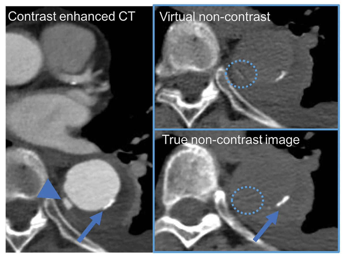
The IQon Spectral CT has had a profound effect on diagnostic accuracy, said Drs. Gupta and Truwit, specifically in CT angiograms of the chest and aorta, where a true noncontrast image is followed by a contrast-enhanced image. Using spectral CT, a virtual noncontrast image can effectively be created by subtracting the iodine from the contrast-enhanced image. Clinicians can confidently diagnose cardiac disease while also reducing the number of scans.
“When you see a regional part of the lung that’s not getting perfusion on the iodine map, it’s pretty obvious that there is going to be a pulmonary embolus at the apex of the oligemic wedge,” said Dr. Truwit. “Whether it’s new or old is another question, but basically, we get to the right answer and then work our way back. When we’re processing the spectral data, highlighting where the perfusion deficit starts and look to the different slices, we’ll see the blood clot jump across the table at us.”
“IQon Spectral CT technology definitely gives me the ability to make a more confident diagnosis,” said Dr. Gupta. “For example in case of atrial appendage occlusion (WATCHMAN) device placement, spectral CT helps in confidently finding the thrombus in the left atrial appendage, which is a contraindication to device placement. If you place a device when there’s a thrombus in situ, that thrombus may dislodge and embolize to the brain and cause stroke and other damage.”
As additional clinical uses for IQon spectral CT technology continue to be proven, even more patients may benefit from more accurate and more timely diagnoses, resulting in better patient care.
Results from case studies are not predictive of results in other cases. Results in other cases may vary.