Duplication of gallbladder and horse-shoe kidney
Images

CASE SUMMARY
A 9-year-old boy was referred to the Department of Radiology with complaints of dull pain in the right upper quadrant of abdomen, nausea, vomiting and feeling of unusual fullness after meals. Physical exam showed no evidence of tenderness in the right upper quad rant and negative Murphy’s sign. Laboratory findings, including transaminases, lactic dehydrogenase, total bilirubin, alkaline phosphatase, and white blood cell count, were within normal limits. The patient’s temperature was 37.5°C and the rest of his vital signs were normal. Ultrasound and contrast-enhanced computed tomography (CECT) of abdomen and magnetic resonance cholangiopancreatography (MRCP) was performed.
IMAGING FINDINGS
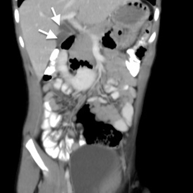
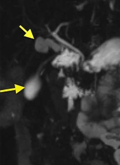
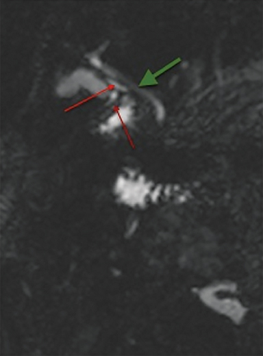
Ultrasound of abdomen showed two anechoic ellipsoid cystic structures in the gallbladder (GB) fossa. Both the kidneys were malrotated with poor visualization of the inferior poles (not shown here). CECT of abdomen was performed. The inferior pole of both the kidneys were medially rotated and fused in the midline anterior to the aorta and inferior vena cava (IVC), with no evidence of hydroureteronephrosis and renal calculi (Figure 1). Two hypodense ellipsoid cystic structures were noted in GB fossa with two cystic ducts joining to form a common duct, which communicates with the common hepatic duct to form the common bile duct. These findings were confirmed on MRCP (Figures 2, 3A, 3B).
DIAGNOSIS
Y-shaped type (vesica fellea duplex) duplication of gallbladder with horseshoe kidney
DISCUSSION
Double gallbladder is a very rare congenital biliary anomaly with a reported incidence of one per 4,000 persons.1 Several anomalies have been associated with gallbladder duplication, including foregut malformations and aberrant hepatic and mesenteric vessels.2 However, no case has been reported of association of duplicated gall bladder with horseshoe kidney to date, which makes this case even rarer. Anatomic duplications of the gallbladder are classified according to Boyden’s into three types: 1) Y-type gallbladder consists of two completely separate gallbladders, each with a cystic duct. The two ducts join to form a common cystic duct before entering the common bile duct; 2) H-type or ductular gallbladder, the cystic and accessory cystic ducts enter the common bile duct separately. This is the most common variant of gallbladder duplication) trabecular gallbladder in which the accessory cystic duct enters the right intrahepatic system.3
It is important to recognize gallbladder duplication due to the clinical, surgical, and imaging difficulties. Accurate pre-operative diagnosis of this anomaly becomes important to prevent possible surgical complications and repeated surgery. Differential diagnosis for duplicate gallbladder includes a folded gallbladder, choledochal cyst, phrygian cap, pericholecystic fluid, gallbladder diverticulum, and vascular band across the gallbladder.
Ultrasound is the first step for evaluation. CT is a simple imaging modality for the diagnosis of double gallbladder. It should be supplemented with MRCP or endoscopic retrograde cholangiopancreatography (ERCP) to detail biliary tract anatomy and its variations.4
When double gallbladder is diagnosed, surgery is recommended. As in our case, surgery was performed and pre-surgical imaging findings were confirmed post-operatively. No intervention is required for incidentally found asymptomatic and uncomplicated horse shoe kidney.
CONCLUSION
It is very important to recognize duplicated gallbladder preoperatively to prevent surgical complications and repeated surgery.
REFERENCES
- Boyden EA. The accessory gallbladder: An embryological and comparative study of aberrant biliary vesicles occurring in man and domestic mammals. Am J Anat. 1926;38:177-231.
- Udelsman R, Sugarbaker PH. Congenital duplication of the gallbladder associated with an anomalous right hepatic artery. AJS. 1985;149:812-815.
- Desolneux G, Mucci S, Lebigot J, Arnaud JP, Hamy A. Duplication of the gallbladder: A case report. Gastroenterol Res Pract. 2009;2009:1-3.
- Gocmen R, Yesilkaya Y. Imaging findings of gallbladder duplication due to two cases: Case report and review of literature. Med Ultrason. 2012;14:358-360.
Citation
K C. Duplication of gallbladder and horse-shoe kidney. Appl Radiol. 2016;(1):34-36.
January 7, 2016