Deep-Learning Model Estimates Breast Density and Cancer Risk
Images
Breast density, defined as the proportion of fibro-glandular tissue within the breast, is often used to assess the risk of developing breast cancer. While various methods are available to estimate this measure, studies have shown that subjective assessments conducted by radiologists based on visual analogue scales are more accurate than any other method.
As expert evaluations of breast density play a crucial role in breast cancer risk assessment, developing image analysis frameworks that can automatically estimate this risk, with the same accuracy as an experienced radiologist, is highly desirable. To this end, researchers led by Professor Susan M Astley from the University of Manchester, United Kingdom, recently developed and tested a new deep learning-based model capable of estimating breast density with high precision. Their findings are published in the Journal of Medical Imaging.
“The advantage of the deep learning-based approach is that it enables automatic feature extraction from the data itself,” explains Astley. “This is appealing for breast density estimations since we do not completely understand why subjective expert judgments outperform other methods.”
Typically, training deep learning models for medical image analysis is a challenging task owing to limited datasets. However, the researchers managed to find a solution to this problem: instead of building the model from the ground up, they used two independent deep learning models that were initially trained on ImageNet, a non-medical imaging dataset with over a million images. This approach, known as “transfer learning,” allowed them to train the models more efficiently with fewer medical imaging data.
Using nearly 160,000 full-field digital mammogram images that were assigned density values on a visual analogue scale by experts (radiologists, advanced practitioner radiographers, and breast physicians) from 39,357 women, the researchers developed a procedure for estimating the density score for each mammogram image. The objective was to take in a mammogram image as input and churn out a density score as output.
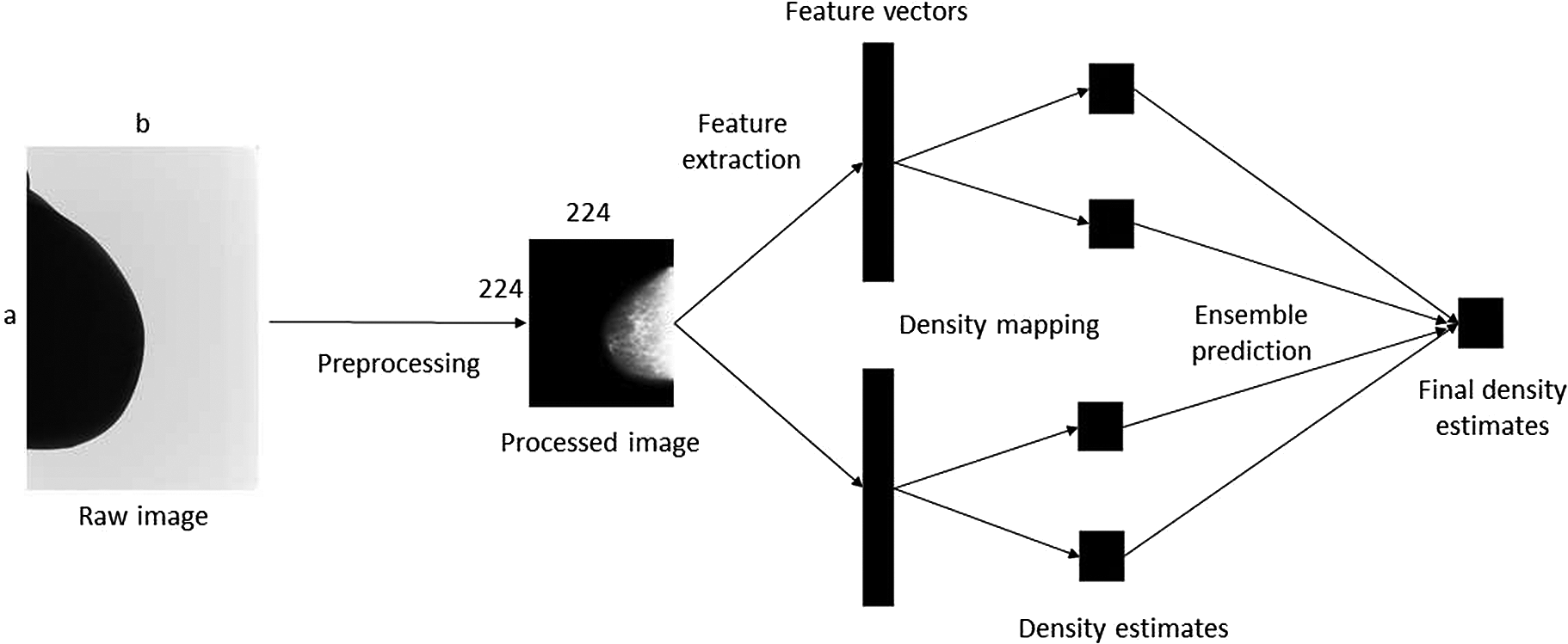
The procedure involved preprocessing the images to make the training process computationally less intensive, extracting features from the processed images with the deep learning models, mapping the features to a set of density scores, and then combining the scores using an ensemble approach to produce a final density estimate.
With this approach, the researchers developed highly accurate models for estimating breast density and its correlation with cancer risk, while conserving the computation time and memory. “The model’s performance is comparable to those of human experts within the bounds of uncertainty,” says Astley. “Moreover, it can be trained much faster and on small datasets or subsets of the large dataset.”
Notably, the deep transfer learning framework is useful not only for estimating breast cancer risk in the absence of a radiologist but also for training other medical imaging models based on its breast tissue density estimations. This, in turn, can enable improved performance in tasks such as cancer risk prediction or image segmentation.