Breast Cancer Risk Assessment Improved by Combining AI Models
Images
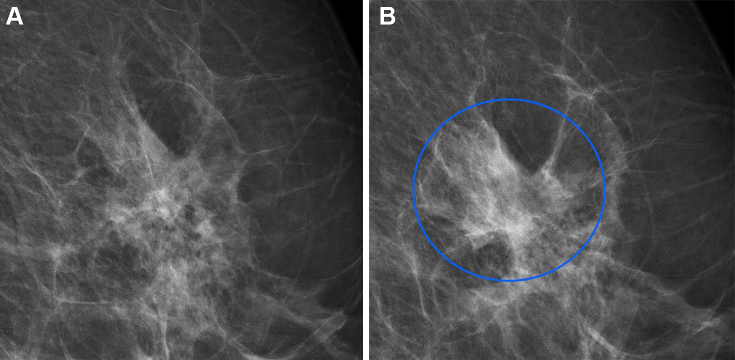
A patient’s cancer risk assessment is improved by combining artificial intelligence (AI) systems for short- and long-term breast cancer risk, according to a study published in Radiology.
Most breast cancer screening programs take a one-size-fits-all approach and follow the same protocols when it comes to determining a woman’s lifetime risk of developing breast cancer. Using mammography-based deep learning models may improve the accuracy of breast cancer risk assessment and can also lead to earlier diagnoses.
“About 1 in 10 women develop breast cancer throughout their lifetime,” said study author Andreas D Lauritzen, PhD, from the Department of Computer Science at the University of Copenhagen in Denmark. “In recent years, AI has been studied for the purpose of diagnosing breast cancer earlier by automatically detecting breast cancers in mammograms and measuring the risk of future breast cancer.”
A variety of AI tools exist to aid in detecting cancer risk. Diagnostic AI models are trained to detect suspicious lesions on mammograms and are well suited to estimate short-term breast cancer risk.
More suitable for long-term breast cancer risk are texture AI models, capable of identifying breast density. Women with dense breast tissue are at higher risk of developing breast cancer and may benefit from supplemental MRI screening.
“It is important to enable reliable and robust assessment of breast cancer risk using information from the screening mammogram,” Dr Lauritzen said.
For this study, Dr Lauritzen and his research team sought to identify whether a commercially available diagnostic AI tool and an AI texture model, trained separately and then subsequently combined, may improve breast cancer risk assessment.
The researchers used the diagnostic AI tool Transpara and a texture model that was developed by the researchers. A Dutch training set of over 39,000 exams was used to train the models. The short- and long-term risk models were combined using a three-layer neural network.
The combined AI model was tested on a study group of more than 119,000 women who were included in a breast cancer screening program in the Capital Region of Denmark between November 2012 and December 2015. The average age of the women was 59 years.
Compared to the diagnostic and texture models alone, the combined AI model showed an overall improved risk assessment for both interval and long-term cancer detection. Interval cancers are those that are found between routine screenings.
The model also enabled identification of women at high risk for breast cancer. Women identified by the combined model as having the 10% highest combined risk accounted for 44.1% of interval cancers and 33.7% of long-term cancers.
Using AI to identify a women’s breast cancer risk from a single mammogram will not only result in earlier cancer detection but can also improve the strain on the health care system due to the worldwide shortage of specialized breast radiologists.
“Current state-of-the-art clinical risk models require multiple tests such as blood work, genetic testing, mammogram and filling out extensive questionnaires, all of which would substantially increase the workload in the screening clinic,” Dr Lauritzen said. “Using our model, risk can be assessed with the same performance as the clinical risk models but within seconds from screening and without introducing overhead in the clinic.”
Related Articles
Citation
Breast Cancer Risk Assessment Improved by Combining AI Models. Appl Radiol.
August 29, 2023