AI Tool May Detect Metastatic Breast Cancer in MRI
Images

Researchers at UT Southwestern Medical Center have developed a novel artificial intelligence (AI) model to improve the detection of breast cancer metastasis, which could reduce the need for needle or surgical biopsies.
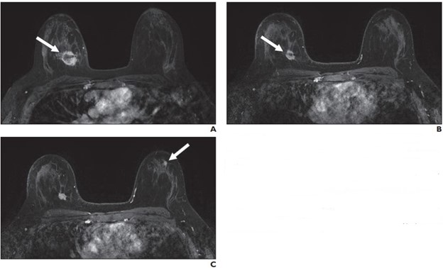
The noninvasive model uses standard magnetic resonance imaging (MRI), paired with machine learning AI, to detect axillary metastasis – the presence of cancer cells in the lymph nodes under the arms.
“Most breast cancer deaths are due to metastatic disease, and the first site is usually an axillary lymph node,” said study leader Basak Dogan, MD, Professor of Radiology, Director of Breast Imaging Research, and member of the Harold C. Simmons Comprehensive Cancer Center at UT Southwestern. “Determining nodal status is critical in guiding treatment decisions, but traditional imaging techniques alone do not have enough sensitivity to rule out axillary metastasis. That often requires patients to undergo invasive procedures that involve radioisotope and dye injection followed by surgery to remove and test whether the axillary nodes harbor cancer cells.”
The research, published in Radiology: Imaging Cancer, showed that the AI model was significantly better at identifying patients with axillary metastasis than MRI or ultrasound. In clinical practice, the AI model would have helped avoid 51% of benign (noncancerous) or unnecessary surgical sentinel node biopsies while correctly detecting 95% of patients with axillary metastasis.
“That’s an important advancement because surgical biopsies have side effects and risks, despite having a low probability of a positive result confirming the presence of cancer cells,” Dr Dogan noted. “Improving our ability to rule out axillary metastasis during a routine MRI – using this model – can reduce that risk while enhancing clinical outcomes.”
The retrospective study used dynamic contrast-enhanced breast MRI exams from 350 newly diagnosed breast cancer patients at UT Southwestern and the Moody Center for Breast Health, which is located on Parkland Health’s main campus in Dallas. All had known nodal status. The images, along with a range of clinical measures, were used to train the AI model to identify axillary metastasis using machine learning techniques.
Because the model is used in conjunction with standard imaging exams, it can also eliminate the stress and expense of additional testing for many patients.
“Patients with benign findings from traditional MRI exams or needle biopsies are often subjected to sentinel lymph node biopsy because those tests can miss a significant proportion of metastasis,” Dr Dogan explained. “Our research demonstrates that it’s possible to identify – with a high degree of accuracy – patients who are nonmetastatic, which benefits the patient and also allows the physician to tailor treatment.”
The research builds on previous studies at UT Southwestern related to breast cancer imaging and the development of predictive tools to detect metastasis.
“Our study is a testament to UT Southwestern’s commitment to impactful research that addresses real-world health care challenges,” Dr Dogan said. “The development and validation of AI models for medical imaging holds great promise in helping us in the fight against breast and other cancers, and this new tool is a significant step forward.”
Researchers are continuing to refine the image analysis process and are looking to include more varied data to validate their findings.