Acute calcific prevertebral tendonitis of the longus colli muscle
Acute calcific tendonitis of the longus colli muscle
Findings
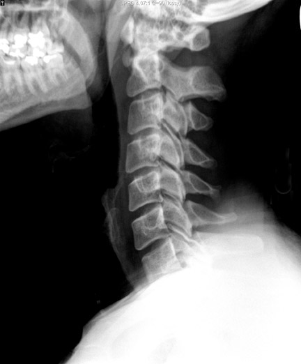
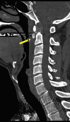
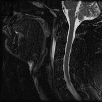
A lateral radiograph of the soft tissues of the neck reveals smooth, mild thickening of the prevertebral soft tissues (5 mm), with a 1.3 cm focus of amorphous calcification anterior to the C2 vertebrae (Figure 1). A contrast-enhanced CT exam (with sagittal reconstructed images) showed a smooth, lenticular-shaped fluid collection within the prevertebral soft tissues. In addition, amorphous calcification was seen embedded within the superior oblique tendon (proximal tendon of the longus colli muscle; Figure 2). An MRI examination also depicted a lenticular shaped prevertebral fluid collection with amorphous calcification in the proximal fibers of longus colli tendon, at its origin from the anterior arch of the C1 vertebrae (Figure 3).
Discussion
Acute retropharyngeal tendonitis (calcific tendonitis of the longus colli), initially described by Hartley in 1964, is an acute inflammation condition of the longus colli tendon, which is related to calcium hydroxyapatite deposition in the superior oblique fibers of the longus colli muscles.1-3 The longus colli muscle extends from the anterior arch C1 to the T3 vertebrae.4 Patients most often present during the third through sixth decades of life5 with acute neck pain, stiffness, odynophagia, low-grade fever and mild leukocytosis.2-8 The clinical presentation can be difficult to differentiate from a retrophayngeal abscess. Radiology can play a key role in the diagnosis: specifically, by identifying characteristic, amorphous calcification in the proximal fibers of the longus colli (just inferior to the anterior arch of theatlas); and, by identifying a prevertebral fluid collection/effusion, which is considered nearly pathognomonic of this entity. The prevertebral fluid collections in this entity tend to be smooth, linear/lenticular and do not contain an enhancing wall (as one may expect with an abscess).3
CONCLUSION
Acute retropharyngeal tendonitis is a cause of acute neck pain that can mimic infection (retropharyngeal abscess, pharyngitis or peritonsilar abscess). Recognition of this entity is critical in order to avoid misdiagnosis and inappropriate treatment with antibiotics and possible"abscess" drainage. In the proper clinical setting, the presence of amorphous calcification anterior to C1/ C2, along with prevertebraledema/fluid, is nearly pathognominic of this entity.
CASE FOLLOW-UP
Given the clinical symptoms, physical examination and specific imaging findings, the diagnosis of acute calcific tendonitis of the longus colli was established. At this point, empiric antibiotic therapy was discontinued, and the patient was treated with nonsteroidalanti-inflammatory medication. The patient responded well to conservative treatment and subsequently made a full recovery.
- Hartley J. Acute cervical pain associated with retropharyngeal calcium deposit: A case report. J Bone Joint Surg Am. 1964;46:1753-1754.