ASU Develops First-of-Its-Kind Compact X-ray Light Source
 Arizona State University has commissioned a first-of-its-kind instrument that will help scientists see deeper into matter and living things. The device, called the compact X-ray light source (CXLS), generated its first X-rays on the night of Feb. 2 by ASU scientists.
“This marks the beginning of a new era of science with compact accelerator-based X‑ray sources,” said Robert Kaindl, PhD, who directs ASU’s Compact X-ray Free Electron Laser (CXFEL) Labs at the Biodesign Institute and is a professor in the Department of Physics. “The CXLS provides hard X-ray pulses with high flux, stability and ultrashort durations, in a very compact footprint. This way, matter can be resolved at its fundamental scales in space and time, enabling new discoveries across many fields — from next-generation materials for computing and information science, to renewable energy, biomolecular dynamics, drug discovery and human health.”
Building the compact X-ray light source is the first phase of a larger CXFEL project, which aims to build two instruments including a coherent X-ray laser. As the first-stage instrument, the ASU CXLS generates a high-flux beam of hard X‑rays, with wavelengths short enough to resolve the atomic structure of complex molecules. Moreover, its output is pulsed at extremely short durations of a few hundred femtoseconds —well below a millionth of one millionth of a second — and thus short enough to directly track the motions of atoms.
Such capabilities have so far only been available at large X-ray free-electron laser (XFEL) facilities, whose number has been limited to only a handful of sites worldwide due to their size and billion-dollar level construction costs. Here, the ASU device provides a uniquely compact facility for ultrashort X-rays that fits into the size of a basement, making leading-edge X-ray science accessible to a university campus.
X-rays have long propelled medical and scientific discoveries far beyond what we can see with the naked eye. Applications transcend the scales, from X-ray astronomy to peer at the universe, to X-ray medical imaging at the human scale, down to resolving complex materials and biomolecules at their atomic scale. Such methods however remained largely static, keeping hidden the ultrarapid dynamics at the heart of proteins at work in our bodies and many other processes in matter.
Only recently researchers have been able to access such dynamics with ultrashort X-ray pulses as generated by XFELs. These ultrashort X-ray pulses can capture atomic-scale molecules akin to how photographers can make a still picture of a hummingbird’s wings via the short bursts of strobe lights used in high-speed photography. Moreover, ultrashort X-rays enable new ways to capture structures of proteins that are otherwise difficult to crystallize.
Here, ASU’s compact X-ray light source will provide key capabilities to help understand both the three-dimensional arrangement and motions of atoms, helping to resolve protein function or advance drug discovery by seeing how a drug interacts with its molecular target, for example.
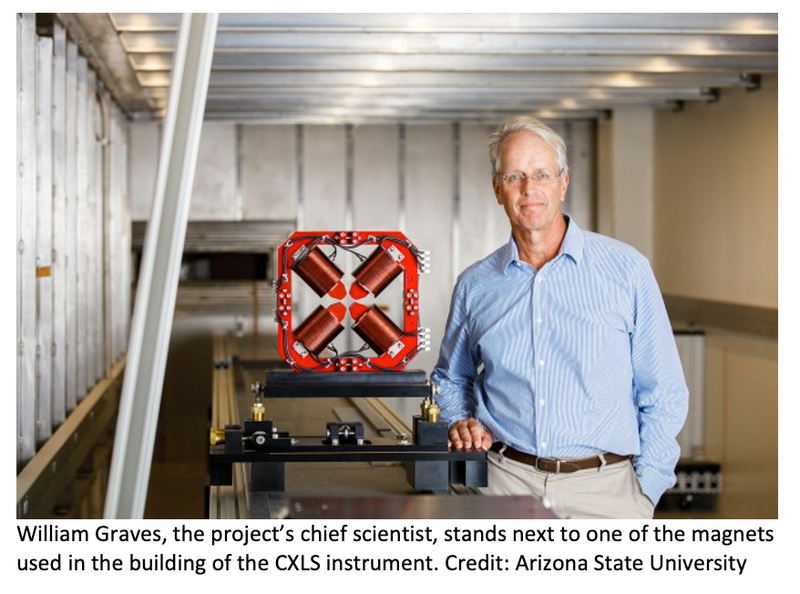
“This is giving us a new tool to look at medical science and semiconductors and all kinds of imaging in different ways,” said William Graves, PhD, the project’s chief scientist, director of accelerator science at the Biodesign Institute and a professor in ASU’s College of Integrative Sciences and Arts. “What this machine allows us to do is see soft tissue changes. We can see blood flowing in blood vessels. We can see individual nerves. We can see down to the cellular level.”
The new ASU CXLS facility includes three main components:
- A table-top particle accelerator that produces a stable electron beam with energies reaching up to 30 million (30 MeV) electron volts.
- A high-powered infrared laser that interacts with the electron beam, in turn producing ultrashort hard X-ray pulses with up to 20 keV photon energy.
- Science experiment chambers and a tunable excitation laser to carry out studies of X-ray interactions with a wide variety of research targets.
To make the first X-rays, the CXLS instrument was powered up to deliver around 4 keV photon energy. The first step takes place in the photoinjector of the light source. There, UV laser pulses are applied to a copper surface — at a rate of 1,000 pulses per second — each releasing a bunch of electrons into vacuum which are then accelerated in a strong electric field. Next, the electron bunches are driven by a linear accelerator to nearly the speed of light and travel through a series of magnets that guide and focus the beam into an interaction chamber.
In the final step, an infrared laser is shot nearly head-on into the path of the oncoming electrons. This results in the emission of powerful X-rays, in a process known as inverse Compton scattering, where the laser is key to the compact facility size. Strong magnetic fields shepherd the electrons into a capture sink. The emitted X-rays are sent downstream to interact with the sample of interest, such as proteins or other molecules (for the first X-ray demonstration, this step was omitted).
The experiment was performed and confirmed by detecting light emission from a YAG scintillator screen. Scientists watched and monitored the beam activities and confirmed the generation of X-rays in their data analyses.
“I am intrigued by technical innovations that greatly shrink the size and cost of these machines,” Dr Graves said. “Turning on this novel light source represents the culmination of a decade of groundwork, from theory to design to build out, all the while overcoming obstacles such as supply-chain delays and other disruptions due to the worldwide pandemic.”